Author: Kathryn Atkins

Trending with Impact: Crosstalk In the Tumor Microenvironment

Authors of this review paper discuss the complex crosstalk between cancer stem cells and macrophages, and potential anti-cancer strategies for future studies.

Figure 1: Main roles of tumor associated macrophages in cancer development and maintenance.
Figure 1: Main roles of tumor associated macrophages in cancer development and maintenance.

The Trending with Impact series highlights Oncotarget publications attracting higher visibility among readers around the world online, in the news, and on social media—beyond normal readership levels. Look for future science news and articles about the latest trending publications here, and at Oncotarget.com.

Listen to an audio version of the article

A review paper published by researchers from the University of Modena and Reggio Emilia in Italy and the Sulaiman AlRajhi Medical School in Saudi Arabia is trending and titled, “Cancer stem cells and macrophages: molecular connections and future perspectives against cancer.” 

“The aim of this review is to define the complex crosstalk between these two cell types and to highlight potential future anti-cancer strategies,” Dr. Beatrice Aramini said, a thoracic surgeon and scientist from the University Hospital of Modena Reggio Emilia.

There have been numerous studies published over recent decades in an effort to understand the molecular mediators of cancer stem cells (CSCs) and tumor associated macrophages (TAMs). Several studies have contributed to bringing light to some of the complex crosstalk that occurs between these two cell types and within the tumor microenvironment. The authors of this review paper reference hundreds of studies and offer a thorough audit and analysis of the current state of this research.

About the Writers of the Review Article

“I mainly focus on lung cancer,” Dr. Aramini said. “I started this project about cancer stem cells in lung cancer since 2017, at University Hospital of Modena Reggio Emilia, joining the laboratory of cell therapies directed by Professor Massimo Dominici with the chief of medical oncology at University Hospital in Modena.”

Dr. Valentina Masciale, co-author, is a research fellow at the University Hospital of Modena Reggio Emilia. Dr. Masciale’s professional experience began by studying missing stroma cells. She then studied stem cells in regenerative medicine and currently she is working with Dr. Aramini on a project focused on lung cancer stem cells. The paper they wrote was revised and approved by seven other contributing authors.

“In this review, we describe the importance of cancer stem cells as the key drivers of cancer initiation and progression due to their unlimited cell renewal capacity and their ability to induce tumor formation,” Dr. Aramini said.

Introduction to Cancer Stem Cells

“Cancer stem cells (CSCs) constitute a cancer cell subpopulation similar to the other stem cell types in terms of self-renewal and multilineage differentiation potential but drive tumor development besides heterogeneity and dissemination of cancer cells [19].”

In 1997, Bonnet and Dick were the first researchers to report the existence of cancer stem cells in the tumor, in acute myeloid leukaemia. Since then, however, a standard marker to identify CSCs still has yet to be found. 

“One of the main obstacles to proving the CSC model is the difficulty in identification and isolation of these cells [73391].”

The authors explain that one of the problems with finding a marker such as this is that many markers found are not only able to detect CSCs, but they also detect non-tumor cells. This represents a major obstacle when developing new therapies to target CSCs only. The authors note that recently there have been several gene markers described by researchers for CSCs in different tumors, including brainbreastblood, and lung.

“Indeed, there are currently no markers able to distinguish between stem cells and CSCs. Thus far, the best markers identified are those of onco-fetal stem cells, which are absent in adult organs and present in cancer cells [4548].”

Theories About the Role of Cancer Stem Cells

This review refers to a few theories about the role of CSCs in cancer progression. One theory is based on the premise that tumor tissue is hierarchically organized into different types of cells, with the CSC subpopulation as the top of this hierarchy. In this theory, the other levels consist of additional differentiated tumor cells or cells with a limited proliferative potential. The “clonal evolution theory” hypothesizes that a rampant mutating cell is the catalyst for tumor progression. 

“Peter Nowell was the first to describe the ‘clonal evolution theory,’ defining cancer as a complex process resulting from the development of a single out-of-control cell with multiple cell mutations that result in the progression of the tumor, which is kept viable through the selection of the most aggressive clones [89].” 

Since the discovery of CSC plasticity and the possibility of switching from stem to non-stem cells, researchers have gained a more complex picture of the origin of tumor heterogeneity and more theories about the role of cancer stem cells in tumor progression.

“An opposing theory is based on the concept that CSCs are a group of cells endowed with a high self-renewal capacity that can set different phenotypes of tumorigenic cells [1888].” 

Cancer Stem Cells and Macrophages

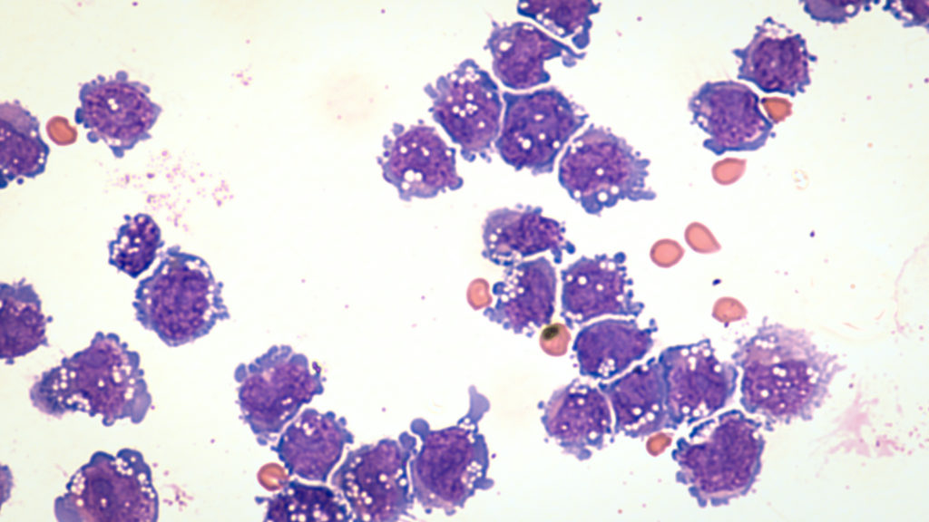
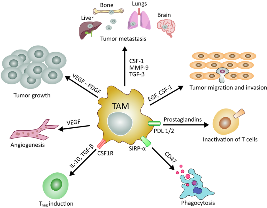
The researchers explain that macrophages are large specialized phagocytic cells that exist in tissues or at infection sites which act as part of the immune system. Arising from the bone marrow, macrophages perform multiple functions and roles in normal and tumor microenvironments, including pro-inflammatory activities and anti-inflammatory processes. Tumor-associated macrophages (TAM) comprise up to 50% of the tumor mass and have a close relationship with CSCs.

“The rising interest on these type of cells comes from recent study demonstrating that high number of tumor-associated macrophages correlate with the poor clinical prognosis in many solid tumors, including lung cancer, which is the field of our research group at the University Hospital of Modena,” Dr. Masciale said. “Another important aspect is the protective role of the tumor-associated macrophages play on tumors undergoing chemotherapy, which may impact the chemotherapy resistance and consequent tumor relapse.”

In recent studies, high numbers of TAMs in lung tumors, gastric cancer, and other cancer types, have been shown to correlate with a poor clinical prognosis. Macrophages are recruited to the tumor and, through crosstalk, provide protection to the tumor, contribute to immunosuppression in the tumor microenvironment and, eventually, drug resistance.

“The primary cause of failure in cancer treatment is the emergence of drug resistance that promotes the tumor spreading,” Dr. Masciale said.

“Cross-talk between CSCs and TAMs involves the recruitment of TAMs through vascularization and the release of chemokines by TAMs to preserve the quiescence of CSCs and modification of their antigens to escape from recruitment by immune cells.”

Future Perspectives

“Although most TAM-targeting strategies are in the pre-clinical stages, several factors used for TAMs depletion have already been tested in clinical trials [271272].”

Current efforts are underway to reprogram or inhibit the tumor-protective properties of tumor associated macrophages. Researchers are also investigating potential strategies to increase the efficacy of chemotherapy through nano-drug delivery to TAMs.

“Due to the significance of the tasks in which TAMs are involved, TAMs are increasingly becoming principal targets of novel therapeutic approaches, especially in the field of nanomedicine.”

The authors believe that targeting TAMs could trigger various reactions in the tumor, which are difficult to predict even given the individual variability from patient to patient. They also explain that targeting TAMs with CSCs offers another potential for treating different tumors to better control cancer progression and avoid tumor dissemination. 

“In summary, generating new information about the interaction between TAMs and CSCs will be one of the most important challenges for the development of more effective targeted cancer therapies.”

Click here to read the full scientific review, published in Oncotarget.

Click here to read or watch an interview with the authors Behind the Study.

Oncotarget is a unique platform designed to house scientific studies in a journal format that is available for anyone to read—without a paywall making access more difficult. This means information that has the potential to benefit our societies from the inside out can be shared with friends, neighbors, colleagues and other researchers, far and wide.

For media inquiries, please contact media@impactjournals.com.

Trending with Impact: Murine Model of Cancer-derived Myocardial Damage

Researchers in this study employed one of the few available murine cachexia models and validated its ability to be used in future studies of cancer-derived myocardial damage.

Part of Figure 2: Alterations in the myocardium of CT26-inoculated BALB/c mice.

The Trending with Impact series highlights Oncotarget publications attracting higher visibility among readers around the world online, in the news, and on social media—beyond normal readership levels. Look for future science news and articles about the latest trending publications here, and at Oncotarget.com.

Listen to an audio version of this article

Cachexia, a complex metabolic syndrome characterized in part by the loss of muscle mass, can account for up to 30% of all cancer-related deaths. Myocardial atrophy, or cardiac remodeling/degradation, is a phenotype of cachexia and a common cause of death.  

“The causes of cancer-derived myocardial impairment might be the effects of cancer itself, background heart disease, and influence of cancer treatments; however, they have not been given much clinical importance, and specific treatment efforts are delayed [8].”

Researchers from Nara Medical University, Hanna Central Hospital, and Hoshida Minami Hospital in Nara and Osaka, Japan, and Nantong University in Jiangsu Province, China, note that while myocardial damage in cancer patients is known to be a cause of death, there are few murine cachexia models available to evaluate cancer-derived heart disorders. Thus, there is a need for further studies that may allow researchers to establish an intervention to prevent myocardial damage in cancer patients.

“In this study, we used the mouse cancer cachexia model that we previously established [14] to examine the status of cancer-derived myocardial impairment reported in literature, and validate our model for studying cancer-derived myocardial impairment.”

The Study

Some causes of cancer-derived myocardial impairment have been reported as cancer-induced cytokines, oxidative stress, depletion of antioxidants, and protein catabolism as a result of AKT/mTOR inhibition.

“Despite these advances in our understanding, the multifactorial mechanisms underlying cancer-derived myocardial impairment remain incompletely understood, necessitating further investigations to elucidate the molecular mechanisms and prevent myocardial damage in cancer patients.”

The researchers previously established a mouse cancer cachexia model. In this study, they aimed to validate their model by employing it in the examination of cancer-derived myocardial impairment that has been reported in previous literature. Their study enlisted the mouse model, CT26 colon cancer cell cultures, protein extraction, histological analysis, immunoblot analysis, enzyme-linked immunosorbent assay (ELISA), mitochondrial stress tests (Seahorse assay), glycolytic stress tests, and statistical analysis. 

Conclusion

“In summary, our established mouse cachexia model showed various myocardial changes associated with cancer cachexia such as oxidative stress in the myocardium, energy metabolism, autophagy, and inflammatory cytokines.”

Results obtained by the researchers in this study using their mouse cachexia model are congruent with previously reported results about cancerous myocardial damage, and therefore provide reasonable evidence that it may be used in future studies.

“The established mouse cachexia model can therefore be considered useful for analyzing cancer-derived myocardial damage.”

Click here to read the full scientific study, published in Oncotarget.

Oncotarget is a unique platform designed to house scientific studies in a journal format that is available for anyone to read—without a paywall making access more difficult. This means information that has the potential to benefit our societies from the inside out can be shared with friends, neighbors, colleagues and other researchers, far and wide.

For media inquiries, please contact media@impactjournals.com.

Trending with Impact: New Prognostic Parameters for Breast Cancer

Different imaging and assessment tools across multiple clinics can result in varied prognostic values. Researchers from Japan conducted a retrospective study of harmonized pretreatment volume-based quantitative FDG-PET/CT parameters for prognostic values in breast cancer patients.

PET Scan image of whole body Comparision Axial, Coronal plane in patient breast cancer recurrence treatment.

The Trending with Impact series highlights Oncotarget publications attracting higher visibility among readers around the world online, in the news, and on social media—beyond normal readership levels. Look for future science news and articles about the latest trending publications here, and at Oncotarget.com.

Listen to an audio version of this article

Breast cancer consists of a wide variety of tumor types, symptoms, disease progression courses, and responses to treatments. In the clinic, researchers decide which disease interventions to use by evaluating the patients’ stage of tumor-node-metastasis (TNM), histologic tumor grade, and the levels of hormone receptors and molecular markers that are present.

Standardized uptake value (SUV), metabolic tumor volume (MTV), and tumor lesion glycolysis (TLG) are derived from 18F-fluorodeoxyglucose positron emission tomography/computed tomography (FDG-PET/CT). These variables have also been reported to correlate with clinicopathological prognostic factors and are considered predictive factors of prognosis.

Breast Cancer Prognostic Parameters

“Recently, noninvasive diagnostic tools have been gaining popularity for prediction of tumor behavior, with magnetic resonance spectroscopy (MRS) and diffusion-weighted imaging (DWI) with magnetic resonance imaging (MRI) reported to provide surrogate imaging biomarkers showing correlations with clinicopathological prognostic factors [23].”

In a multi-institutional retrospective study in Japan, researchers—from the Hyogo College of Medicine, Nippon Medical School Hospital, National Cancer Center Hospital, Kinki University Faculty of Medicine, and Gunma Prefectural College of Health Sciences—explain that the factors and algorithms used by different assessment tools across multiple clinics can result in varied standardized uptake values. These inconsistencies have provided an opportunity for the researchers to standardize parameters of prognostic values when imaging breast cancer patients to improve patient outcomes.

“Thus, a harmonization strategy is necessary for comparing semi-quantitative PET parameters among available imaging methods, which is a notably relevant issue for multicenter trials employing different PET systems.”

The Study

Researchers gathered records of 546 patients treated from 2010 to 2016 with stage I–III invasive breast cancer. Of those patients, 344 were estrogen receptor (ER)-positive/human epidermal growth factor receptor two (HER2)-negative, 110 were HER2-positive, and 92 were triple-negative. The patients were treated at four separate institutions using different PET/CT scanner systems. In addition to surgeries, chemotherapy, and radiotherapy, patients were assessed during their follow-up appointments.

“Mammography, ultrasound, CT, bone scanning, and FDG-PET/CT were used for determining disease recurrence, metastasis, and progression during follow-up.”

Researchers in this study retrospectively performed histological and statistical analyses of overall survival and recurrence-free survival in patients of each breast cancer subtype group.

“An experienced reader (12 years of experience with oncologic FDG-PET/CT) who had no knowledge of other imaging results or clinical and histopathologic data retrospectively reviewed all of the FDG-PET/CT images.”

They found that the average maximum standardized uptake values (SUVmax) for HER2-positive and triple-negative tumor patients were higher than in patients with ER-positive/HER2-negative tumors.

“Harmonized primary tumor and nodal maximum SUVmax, metabolic tumor volume (MTV), and TLG indicated in pretreatment FDG-PET/CT results were analyzed.”

Conclusion

Results from this study suggest that harmonized PET classifications with final clinical response assessments demonstrate a better ability to predict disease-free survival compared to non-harmonized PET classification.

“We concluded that harmonized quantitative volume-based values, especially those for the primary tumor and nodal SUVmax and TLG, obtained with FDG-PET/CT can provide useful information regarding prognosis for both recurrence and death in patients with operable invasive breast cancer, including all three main subtypes. The findings presented here are considered useful for improving care of individual patients.”

Click here to read the full retrospective study, published in Oncotarget.

Oncotarget is a unique platform designed to house scientific studies in a journal format that is available for anyone to read—without a paywall making access more difficult. This means information that has the potential to benefit our societies from the inside out can be shared with friends, neighbors, colleagues and other researchers, far and wide.

For media inquiries, please contact media@impactjournals.com.

An Overview of Cannabis and Cancer

Researchers review different varieties of cannabinoids, the signaling pathways they affect, and their role in different types of cancer.

Close up of female Cannabis flower with a high production of cannabinoid resin

The Top-Performer series highlights research literature published in Oncotarget that has generated a high Altmetric score. Altmetric scores, located at the top-left of trending Oncotarget papers, provide an at-a-glance indication of the volume and type of online attention the research has received. Read Oncotarget’s Top 100 Altmetric papers.

Listen to an audio version of this article

In a high-rated paper published in 2014 in Oncotarget, researchers from India’s Sanjay Gandhi Post Graduate Institute of Medical Sciences and the United States’ Ohio State University reviewed cannabinoids, their role in different types of cancer, and the signaling pathways they affect. Today, this paper currently presents with an Altmetric Attention score of 200.

“In this review article, we will focus on a broad range of cannabinoids, their receptor dependent and receptor independent functional roles against various cancer types with respect to growth, metastasis, energy metabolism, immune environment, stemness and future perspectives in exploring new possible therapeutic opportunities.”

Cannabinoids and Receptors

“[The] Cannabis sativa plant has been used for several hundreds of years both recreationally and medicinally.”

Researchers trace the earliest archaeological evidence of cannabis medical use back to ancient China, during the Han Dynasty. The use of this plant was recommended for rheumatic pain, constipation, disorders of the female reproductive tract, and malaria, among other conditions. Cannabis sativa contains three major classes of bioactive molecules; flavonoids, terpenoids, and 100+ types of cannabinoids. 

Cannabinoids are a family of complex chemicals that activate and bind to two receptors in mammals named central cannabinoid receptor one (CB1) and peripheral cannabinoid receptor two (CB2). These receptors are found abundantly throughout the central nervous system and immune system.

“CB1/2 receptors are also responsible for proliferation, motility, invasion, adhesion and apoptosis of cancer cells both in vitro and in vivo.”

CB1 and CB2 receptors have been used as targets for the treatment of various diseases, including neurodegenerative diseases such as Alzheimer’s, Parkinson’s, and Huntington’s disease, neuropathic and inflammatory pain, glaucoma, multiple sclerosis, cardiovascular disorders, obesity, and more. Today, in addition to inhibiting nausea and emesis, stimulating appetite, improving mood, and relieving pain and insomnia that cancer patients face, cannabinoids used in the targeted killing of tumor cells has been a major discovery in cancer treatment. 

“In this review article we focused on the role of cannabinoids in different cancer types and the respective signaling pathways.”

Endocannabinoids

“Endogenous cannabinoids which are produced in our body include lipid molecules containing long-chain polyunsaturated fatty acids, amides, esters and ethers that bind to CB1 or CB2 receptors.”

Endocannabinoids act primarily as neuromodulators, or reverse messengers, which can affect the release of neurotransmitters. They also play important role in regulating inflammation, insulin, and fat and energy metabolism, which affects our mood, appetite, pain sensation, inflammation response, and memory. 

Phytocannabinoids

“Phytocannabinoids are only known to occur naturally in significant quantities in the cannabis plant, and are concentrated in a viscous resin that is produced in glandular structures known as trichomes.”

Over 120 phytocannabinoids are capable of interacting within the body’s own biological systems because their structures and behaviors mimic those of endocannabinoids (cannabinoids that are synthesized by our own bodies). The most prevalent natural cannabinoids are delta-9-tetrahydrocannabinol (∆9-THC), cannabidiol (CBD), and cannabinol (CBN). 

Synthetic Cannabinoids

“Synthetic cannabinoids are classified on the basis of chemical structure of molecules and they are capable of a more selective activation of cannabinoid receptors [28].”

The researchers explain that synthetic cannabinoids have been used extensively in pharmacology to gain better insight about their action in order to evaluate the potential use of cannabinoids clinically. 

Within the synthetic category, classical cannabinoids are compounds isolated from the Cannabis sativa plant or its synthetic analogs. Nonclassical cannabinoids “are a family of AC-bicyclic and ACD-tricyclic cannabinoid analogs.” Aminoalkylindoles are non-cannabinoid molecules given cannabis-mimicking capabilities. Eicosanoids are compounds that can enhance or inhibit physiological and pathophysiological responses. These lipid mediators also have an affinity for CB1 and CB2 receptors.

Cannabinoids in Cancer

Multiple studies have shown that THC, CBD, and synthetic cannabinoids can inhibit breast cancer cell proliferation and drive them toward apoptosis.

“It [breast cancer] is classified into three main subtypes according to their molecular profiles: hormone receptor-positive, HER2-positive (ErbB2-positive, a member of EGFR family) and triple-negative tumors [42-43]. Cannabinoid-based medicines have been useful for the treatment of these three breast cancer subtypes.”

In prostate cancer, CB1 and CB2 expression levels are often higher in prostate cancer tissues and several cell lines compared to normal prostate epithelial cells. Studies have found that cannabinoids have either induced cell death or activated pathways that lead to growth inhibition and increased patient survival.

Preclinical cancer models have shown that cannabinoids can alter gene expression, block enzymes, inhibit signaling pathways, and induce apoptosis in mice with lung cancer. In skin and pancreatic cancers, researchers have found that the activation of CB1/2 receptors induced the apoptotic death of tumorigenic cells, without affecting the normal cells. In bone cancer studies, researchers found that cannabinoids reduced pain and bone loss in mice.

“Cannabinoids could halt tumor development without side effects via specific targeting of CB1/CB2 receptor.”

Cannabinoids have anti-tumorigenic properties in glioma, lymphoma, oral cancers, and thyroid carcinoma. In young people, marijuana smoking has been found to increase the incidence of head and neck cancer, however, cannabinoids have anti-tumor properties.

Conclusion

“Cannabinoids exert a direct anti-proliferative effect on tumors of different origin.”

Given that cannabinoid receptors are often demonstrated to be expressed higher in tumor cells than in normal cells, cannabinoids are more specific to cancer cells than to normal cells. The researchers conclude their review by noting that it is important to identify which cannabinoids are most compatible with an individual cancer or disorder to have the greatest impact on patient outcome.

“It is important to understand which of the cannabinoid receptors are expressed and activated in different tumors as each receptor follows a different signaling mechanism.”

Years after this paper was published, subsequent studies have confirmed and expanded on many ideas mentioned in this review, including the regenerative and pharmacological effects of THC, synthetic cannabis used to treat thrombosis, increased expression of CB2 potentially linked to colon cancer, experiments with cannabis extracts, synergistic combinations of cannabinoids, and much more. 

Click here to read the full scientific review, published in Oncotarget.

Oncotarget is a unique platform designed to house scientific studies in a journal format that is available for anyone to read—without a paywall making access more difficult. This means information that has the potential to benefit our societies from the inside out can be shared with friends, neighbors, colleagues and other researchers, far and wide.

For media inquiries, please contact media@impactjournals.com.

Trending with Impact: Novel MicroRNA Underexpressed in Lung Cancer

In search of new ways to target lung cancer cells, researchers in this study demonstrated that miR-708 has anti-tumorigenic properties.

Photomicrograph of fine needle aspiration (FNA) cytology of a pulmonary (lung) nodule showing adenocarcinoma, a type of non small cell carcinoma.

The Trending with Impact series highlights Oncotarget publications attracting higher visibility among readers around the world online, in the news, and on social media—beyond normal readership levels. Look for future science news and articles about the latest trending publications here, and at Oncotarget.com.

Despite the innumerable biomedical advancements made in the detection, classification, and treatment of cancer since the 1971 National Cancer Act, lung cancer survival rates are still staggeringly low. In addition, every year over $12.1 billion is spent on lung cancer care in the United States. Non-small cell lung cancer (NSCLC) contributes to 85% of lung cancers and within this classification there are two main subtypes: adenocarcinoma (LUAD) and squamous cell carcinoma (LUSC).

“Although tumors are differentiated by subtype, LUAD and LUSC are generally treated with the same chemotherapeutics.”

Researchers, from the New Jersey Medical School’s academic health center, Rutgers Biomedical & Health Sciences, say that discovering new biomarkers that can help better distinguish between NSCLC subtypes is necessary to improve patient outcomes. In 2020, they conducted a study of a microRNA that is dysregulated in lung cancer, miR-708, to clarify its tumor suppressive or oncogenic functions within lung cancer cells.

“Lung cancer is a complex collection of deadly diseases that are generally hard to detect and treat. Therefore, it is crucial to develop novel methods to identify, distinguish, and treat lung cancer.”

The Study

The researchers in this study explain that it is crucial to take the entire tumor microenvironment (TME) into consideration when devising treatments for cancerous tumors. Historically, many chemotherapies that have been developed are successful in targeting tumors, but contribute to damaging the surrounding cells and tissues in the TME—contributing to harm and extending recovery time. In newer treatments being developed, researchers have considered the benefits of targeting the pro-tumor effects of particular immune cells and activating the immune system to attack cancer cells.

“miR-708 has previously been described as being both oncogenic and tumor suppressive in lung cancer [63–65]. Therefore, we aimed to clarify the tumor suppressive or oncogenic functions of miR-708 in lung cancer cells.”

This new potential microRNA with potent anti-tumorigenic effects for non-small cell lung cancer (NSCLC) was identified by the researchers. To determine the clinical relevance of miR-708 in lung cancer patients, the researchers analyzed data from The Cancer Genome Atlas (TCGA) using the TCGA-assembler 2 R software package. They used mammalian cell cultures to perform miRNA and 5-Azacytidine treatments, RNA isolation using TRIzol, quantitative real-time RT-PCR, western blot analysis, plasmids, luciferase reporter assays, Enzyme-Linked Immunosorbent Assay (ELISA) analysis, phenotypic assays; Water Soluble Tetrazolium Salts (WST)-1 assay; Ki-67 staining; Annexin V staining; Cell migration assay, and Bioinformatic and statistical analyses.

“We next examined expression of miR-708 in normal and lung cancer cells to determine if our cell lines faithfully replicated clinical data.”

Results

The researchers discovered miR-708 was underexpressed in lung cancer cells compared to normal lung cells. A lower expression of miR-708 correlated with decreased survival in patients with squamous cell carcinoma non-small cell lung cancer. They demonstrated that miR-708 suppressed the production of the pro-tumorigenic hormone called prostaglandin E2 (PGE2) (located in the arachidonic acid (AA) metabolic inflammatory pathway), by directly repressing the expression of COX-2 and mPGES-1 in lung cancer cells.

“We also demonstrated that miR-708 decreases lung cancer cell metabolism (Figure 5), proliferation (Figure 6), survival (Figure 7), and migration (Figure 8).” 

Conclusion

The researchers were left with some outstanding questions about miR-708. First, they wondered why miR-708 expression is decreased in lung cancer cells compared to normal cells in the lungs. They suggest the cause may be the hypermethylation of the ODZ4 promoter region in lung cancer cells, a loss of tumor suppressive transcription factors, repressed CHOP activity, or specifically, the glucocorticoid receptor-alpha (GRα) repression of CHOP activity.

“Our work has identified novel tumor suppressive miR-708 functions by suppressing oncogenic PGE2 production through targeting of COX-2 and mPGES-1. These findings could be the foundation for identifying novel miR-708 targets, as well as regulators of miR-708 expression in cancer.”

“Moreover, our study highlights the need to better understand lung cancer biology to improve diagnosis and treatment of lung cancer, ultimately aiming to increase positive patient outcomes.”

Click here to read the full scientific study, published in Oncotarget.

Oncotarget is a unique platform designed to house scientific studies in a journal format that is available for anyone to read—without a paywall making access more difficult. This means information that has the potential to benefit our societies from the inside out can be shared with friends, neighbors, colleagues, and other researchers, far and wide.

For media inquiries, please contact media@impactjournals.com.

Are Anti-aging Drugs the Key to Cancer Prevention?

In his recent paper, Dr. Mikhail Blagosklonny explains his perspective on the current landscape of anti-aging drug studies, a key differentiation between healthspan and lifespan variables, and the next steps for human use of anti-aging drugs—beyond clinical trials.

Aging in humans seems as natural as aging in leaves—but is it necessary?
Listen to an audio version of this article

The process of human aging is a fascinating mystery. Despite all that we do not know, a handful of researchers have dedicated recent decades to the exciting beginnings of solving this biological riddle. One such researcher is Dr. Mikhail Blagosklonny. As a professor of oncology at the Roswell Park Cancer Institute in Buffalo, New York, and Editor-in-Chief at the AgingandOncotarget journals, Dr. Blagosklonny’s mission is to prevent cancer (and other age-related diseases) by inhibiting the aging process—preventing cancer by maintaining youth.

The cover paper chosen for Oncotarget’s Volume 12, Issue #3, is titled, “The goal of geroscience is life extension;” a research perspective written by Dr. Blagosklonny. In this compelling paper, he reflects on the history of anti-aging studies, the differences between drugs that enhance healthspan versus lifespan, and next steps in the human application of anti-aging drugs. 

Hyperfunction Theory of Aging

“According to the geroscience hypothesis, aging is a risk factor for diseases [127]. According to hyperfunction theory, in contrast, aging is a sum of all age-related diseases, not their risk factors.”

Dr. Blagosklonny defines aging as a continuation of human development, driven partially by growth-promoting pathways which drive age-related diseases—he has coined this as the hyperfunction theory.

“Hyperfunction (inappropriate activation) of these signaling pathways directly drive all age-related diseases, which are manifestations of aging. We just need clinically available inhibitors (drugs) of these signaling pathways to extend both healthspan and lifespan, by slowing aging.”

Increasing Lifespan via Increasing Healthspan

Before beginning his interpretation of data from previous anti-aging research studies, Dr. Blagosklonny emphasises the importance of correctly measuring healthspan and lifespan. As indicated in his paper title, the goal of geroscience is to extend lifespan by way of extending overall healthspan.

“Healthspan is a period of life without age-related diseases [27]. It is disease-free survival.”

Healthspan can be difficult to measure due to the nature and hidden course of many diseases. If one particular disease is subdued by treatment in a study and healthspan appears to be increased (through one marker of health or another), this does not guarantee that other age-related diseases have been nullified by this treatment. Dr. Blagosklonny explains that accurate measurements of healthspan are important because, based on the hyperfunction theory, aging is the sum of all age-related diseases.

“After all, aging is an exponential increase of death with age and should be measured by deadly diseases.”

Another point he makes is that many anti-aging drug trials have presented results finding increased healthspan in mice without demonstrating an increase in lifespan. Given that increased healthspan should always lead to increased lifespan, it is not sufficient to only measure healthspan without measuring lifespan in animal studies of anti-aging drugs. If lifespan is not increased, the drug does not demonstrate longevity or anti-aging properties.

“So how is it possible that some senolytics, NAD boosters and resveratrol, increase healthspan without lifespan? The simplest explanation is that they do not increase healthspan at all, because such studies use irrelevant or ambiguous markers of health.”

Over the years, numerous initially promising anti-aging drugs have been tested and debunked by researchers. No compound has continued to withstand the many tests, or has delivered consistent results, quite like the unique bacterium, rapamycin.

Anti-aging Properties in Rapamycin

Rapamycin was discovered in 1964 in a test tube sample of dirt taken from Easter Island—a highly remote volcanic island in the Pacific ocean, west of Chile. Initially looking for antibiotics (often uncovered in the dirt) researchers found the rapamycin bacteria unexpectedly. To their surprise, this new bacteria created a defensive chemical with the ability to affect the activity of a protein and homeostatic ATP sensor called the mammalian target of rapamycin, or mTOR. mTOR is now known to function in regulatory pathways that are responsible for governing cell growth. 

“It was predicted that rapamycin must extend lifespan before it was shown in any animal [105].”

In 1999, rapamycin was FDA approved to regulate hyperimmunity in transplant patients to help enable their immune system to accept a new organ. Since then, rapamycin’s ability to slow cell growth and proliferation has been widely accepted as an anticancer agent and the focus of anti-aging studies in a number of mouse-modeled trials.

“Since 2009, dozens of studies have shown that rapamycin extends medium and maximum lifespan in both males and females in all strains of normal mice tested, as well as in some cancer-prone and short-lived mice [364070].

Other Drugs With and Without Anti-aging Potential

In this paper, Dr. Blagosklonny categorizes a list of seemingly debunked anti-aging drugs with little or no results, including antioxidants, resveratrol, curcumin, quercetin (used alone), and spermidine. He explains that some of these drugs may have potential when used in combination with other drugs in future studies.

He acknowledges potential in berberine (one study found promising initial results), fisetin (clinically available and safe for human use), 17-alpha-estradiol (only results in male mice thus far), acarbose (blocks digestion of complex carbs), enalapril (decreases oxidative damage), losartan (angiotensin receptor blocker), quercetin with dasatinib (clinically available and safe for human use), and metformin. 

“Some life-extending drugs are already approved for human use: supplements (fisetin, vitamin B3 and its analogs), over-the-counter medicine (aspirin) and prescription drugs (rapamycin, metformin, dasatinib, enilopril).”

Dr. Blagosklonny recalls a famous study of metformin where, at a low doses, it increased lifespan in male mice and, at high doses, it ironically decreased lifespan. Metformin was also tested with rapamycin in this study and demonstrated improved results in extending lifespan.

“Yet, a combination of metformin and rapamycin should be re-tested to include a rapamycin-alone group.”

Conclusion

“I expect that a combination of low doses of pan-mTOR and MEK inhibitors with high doses of rapamycin would extend life further compared with rapamycin alone. That could be the next important advance in the anti-aging field since the discovery of anti-aging properties of rapamycin.”

Dr. Blagosklonny believes that researchers should not wait for the lifespan results of clinical trials in humans to begin widespread application of these drugs, since studies already safely display increased lifespan and longevity in mouse models. He is so convinced by rapamycin that Dr. Blagosklonny is currently taking 10 milligrams of rapamycin per week along with his personalized treatment plan, a ketogenic diet, and exercise to jumpstart the next phase of human anti-aging trials within our lifetime. He notes that medical doctors interested in this topic may email Blagosklonny@rapalogs.com or follow him on Twitter @Blagosklonny.

“This article does not represent medical advice or recommendations to patients. The media should exercise caution and seek expert medical advice for interpretation when referring to this article.” 

Click here to read the full research perspective on Oncotarget.com.

Oncotargetis a unique platform designed to house scientific studies in a journal format that is available for anyone to read—without a paywall making access more difficult. This means information that has the potential to benefit our societies from the inside out can be shared with friends, neighbors, colleagues, and other researchers, far and wide.

For media inquiries, please contact media@impactjournals.com.

Trending with Impact: New Plant Extracts Reveal Anti-aging Properties

In search of natural compounds with previously unknown geroprotective properties, researchers used a strain of budding yeast to test 53 plant extracts for their ability to impact the biology of aging and age-related diseases.

Scientist is sampling a chemical extract from organic natural, research and develop background. Scientific concept is sample project about herbal medicine.

The Trending with Impact series highlights Oncotarget publications attracting higher visibility among readers around the world online, in the news, and on social media—beyond normal readership levels. Look for future science news and articles about the latest trending publications here, and at Oncotarget.com.

As we age, humans are subjected to a wide variety of age-related diseases, such as arthritis, diabetes, heart disease, kidney disease, liver dysfunction, sarcopenia, stroke, neurodegenerative diseases, and many forms of cancer.

Plant extracts have been consumed for hundreds of years in dietary customs and used as traditional herbal medicines in China and in the Mediterranean. Some of these plant extracts are classified by government health agencies, such as Health Canada, as not only safe for human consumption but also as health-improving supplements with clinically proven benefits to human health. Researchers hypothesized that some of these plant extracts (PEs) may have geroprotective properties. A geroprotector is any compound capable of modulating the root cause of aging and age-related diseases to prolong lifespan in modeled organisms and animals. A couple of well-known potential geroprotectors include melatonin and metformin.

In their previous 2016 study, researchers from Concordia University and Idunn Technologies—both located in Quebec, Canada—screened thirty-five plant extracts and identified 6 as capable of prolonging the length of time a cell can survive, or its chronological lifespan (CLS), and delaying chronological aging in the wild type strain of Saccharomyces cerevisiae budding yeast. On a mission to uncover a new set of plant extracts with geroprotectivity, these same researchers conducted a larger screening of plant extracts in a 2020 study. 

“The objective of the present study was to search for previously unknown aging-delaying (geroprotective) PEs. To attain this objective, we conducted a new screen of many extracts from plants used in traditional Chinese and other herbal medicines or the Mediterranean and other diets.”

The Study

In this study, to learn more about new PEs and the mechanisms of aging and longevity, the researchers continued using the wild type strain of Saccharomyces cerevisiae budding yeast. They explained that S. cerevisiae has short and easily measurable replicative (number of times a cell can divide prior to senescence) and chronological lifespans, is completely sequenced, commercially available, and conducive to comprehensive molecular analyses.

Researchers tested 53 new plant extracts on chronologically aging S. cerevisiae budding yeast. The plant extracts were derived from fruits, berries, beans, herbs, flowers, roots, seeds, leaves, stems, whole plants, bulbs, buds, bark, skins, resin, aerial parts, mushroom bodies, and fermented rice.

“In a quest for previously unknown geroprotective natural chemicals, we used a robust cell viability assay to search for commercially available plant extracts that can substantially prolong the chronological lifespan of budding yeast.”

To determine geroprotectivity from these plant extracts, the researchers cultured, diluted, and fed the budding yeast with glucose. Then, after adding the new PEs, they performed a variety of tests and calculated measurements, including: chronological lifespan assay; oxygen consumption assay; plating assay; quantitative assay; fluorescence microscopy; measurements of the frequencies of spontaneous mutations; glucose concentration measurement assay; age-specific mortality rates; the Gompertz slope; the mortality rate coefficient; and mortality rate doubling time.

Results

“We discovered fifteen PEs that extend the longevity of chronologically aging budding yeast.”

The team was able to identify 15 new geroprotective PEs that have not previously been known for their ability to prolong the lifespan of yeast or other organisms. Based on the results of their measurements and assays, the researchers also identified the cellular processes that these PEs engaged in to prolong the yeast’s chronological lifespan.

“Our study provides evidence that each of the fifteen longevity-extending PEs satisfies all the criteria previously proposed for a CRM.”

CR stands for caloric restriction and CRM stands for caloric restriction mimetics. This means that these new PEs were found capable of mimicking the substantial anti-aging effects that calorie restriction has on organisms and animals, without a reduction in calorie intake.

“Each of the fifteen PEs extends the longevity of chronologically aging yeast under non-CR conditions on 2% (w/v) glucose significantly more efficiently than it does under CR conditions on 0.5% (w/v) glucose.”

They found that the PEs extended the longevity of chronologically aging yeast by decreasing the rate of aging, stimulating a hormetic stress response, intensifying mitochondrial respiration, altering the pattern of age-related changes in intracellular reactive oxygen species, and increasing cell resistance to long-term oxidative and thermal stresses.

“Each of the fifteen geroprotective PEs decreases the extent of age-related oxidative damage to cellular proteins, and many of them slow the aging-associated buildup of oxidatively impaired membrane lipids as well as mitochondrial and nuclear DNA.”

In addition to many more findings, the effects of 15 PEs were found to decrease the frequency of mitochondrial DNA mutations in rib2 and rib3 proteins under non-calorie restricted conditions in S. cerevisiae.

Conclusion

The 15 plant extracts in this study that were newly discovered as geroprotective are as follows: berry extract from a small palm commonly known as Saw Palmetto, extract of the aerial parts from a flowering plant commonly known as the St. John’s Wort, extract from the leaf of Yerba Mate, whole plant extract of Yerba Mate, extract from the leaf of Holy Basil Tulsi, extract from the herb of the perennial plant Solidago Virgaurea, Orange fruit extract, whole plant extract from the common Hop (used in beer), Grape skin extract, whole plant extract from the Green Chiretta, root extract from the perennial Goldenseal herb, Fenugreek seed extract, Barberry root bark extract, extract from the leaf, flower, and stem of the common Hawthorn, and leaf extract from the Red-seeded Dandelion.

“Therefore, we are interested in investigating how different combinations of the fifteen geroprotective PEs described here influence the extent of yeast chronological aging delay. We will be looking for the combinations of geroprotective PEs that exhibit synergistic or additive effects on the extent of yeast chronological aging delay.”

Click here to read the full scientific study, published in Oncotarget.

Oncotarget is a unique platform designed to house scientific studies in a journal format that is available for anyone to read—without a paywall making access more difficult. This means information that has the potential to benefit our societies from the inside out can be shared with friends, neighbors, colleagues, and other researchers, far and wide.

For media inquiries, please contact media@impactjournals.com.

Trending with Impact: Bacterial Therapy Experiments in Prostate Cancer

Researchers reveal their positive findings from a study of bacterial cancer therapy using a strain of Salmonella typhimurium in mouse-modeled prostate cancer.

PC-3 human prostate cancer cells stained with blue Coomassie, under a differential interference contrast microscope. - Image
PC-3 human prostate cancer cells stained with blue Coomassie, under a differential interference contrast microscope.

The Trending with Impact series highlights Oncotarget publications attracting higher visibility among readers around the world online, in the news, and on social media—beyond normal readership levels. Look for future science news and articles about the latest trending publications here, and at Oncotarget.com.

Listen to an audio version of this article

Over the past few decades, numerous studies have emerged using the promising strategy of bacteria as vehicles to deliver drugs or genes in tumor‐targeted therapies. Researchers say that bacterial cancer therapy may be able to overcome some of the limitations that conventional cancer therapy is stunted by, including the development of drug resistance. 

Researchers in this study—from Yale University in Connecticut, the University of Missouri, the Harry S. Truman Memorial Veterans Hospital, and the Cancer Research Center in Missouri, and DeSales University in Pennsylvania, U.S.—used a Salmonella typhimurium strain (CRC2631) of bacteria (previously reported to have tumor-targeting capabilities) in prostate cancer-positive mouse-models and evaluated its toxicity, targeting ability, and genetic stability.

“Here, we report the toxicological and in vivo tumor-targeting profiles of CRC2631 in the syngeneic and autochthonous mouse model of aggressive prostate cancer, TRAMP (Transgenic Adenocarcinoma of Mouse Prostate).”

“The B6FVB TRAMP model recapitulates some of the key genetic aspects of human prostate cancer.”

The Study

“VNP20009 is considered as the safety benchmark in bacterial cancer therapy development because it has been safely administered in human cancer patients [7, 30].”

“To determine the safety profile of CRC2631, we performed CRC2631 and VNP20009 comparative toxicological studies in TRAMP animals.”

The team focused on measuring toxicity through treatment-related weight loss and lethality. Groups of 14-week-old B6FVB TRAMP-positive mice were scanned using magnetic resonance imaging. Four mice were dosed with CRC2631, and four were dosed with VNP20009; both treated four times per week. Mice were weighed and monitored daily for four weeks.

Since the CRC2631 bacteria are cleared out through the liver, the researchers also sought to establish the impact of CRC2631 on liver pathology in this bacterial cancer therapy. Two groups of 31-week-old B6FVB TRAMP-positive mice were observed, one treated with four doses of CRC2631 and the other with saline (the control group) at three-day intervals. They used histological staining in the liver to observe differences in necrosis, inflammation, and extramedullary hematopoiesis between CRC2631 and the control group. The team then tested for lethality and the maximum tolerated dose of CRC2631.

“Next, we sought to determine the in vivo tumor-targeting capability of CRC2631 in TRAMP animals.”

Using fluorescence imaging and a chloramphenicol resistance cassette, researchers were able to observe the biodistribution of CRC2631 to determine its tumor-targeting capability in TRAMP-positive mice. Since they knew that CRC2631 is filtered through the liver and that enriched colonies may be found here, researchers used the liver as a way to compare the bacterial load in tumor tissues.

The researchers also tested CRC2631’s genetic stability by gauging its likelihood of regaining toxicity and/or losing tumor targeting capability by performing longitudinal, whole genome sequencing and short nucleotide polymorphism analyses.

“To determine the genetic stability of CRC2631 inside the host, we performed longitudinal whole genome sequencing and short nucleotide polymorphism (SNP) analyses of CRC2631 prior to treatment and tumor-passaged CRC2631 in B6 TRAMP (+) mice.”

In vitro, CRC2631 directly kills prostate cancer cells, however, in vivo, it does not lead to decreased tumor burden. The researchers believe this may be due to the effects of some kind of resistance mechanism in vivo, and tested a combined treatment method of CRC2631 and Invivomab—a checkpoint blockade—in the mouse model.

“CRC2631 targets and directly kills murine and human prostate cancer cells in vitro (Supplementary Figure 2), raising the possibility that unknown resistance mechanisms protect tumor cells from CRC2631-mediated cell death in vivo.”

Results

Researchers explain that in the first two weeks of the study, mice treated with CRC2631 and VNP20009 lost a comparable amount of weight. However, in the second half of the study, VNP20009-treated animals lost progressively more weight than those treated with CRC2631. This revealed that CRC2631 is less toxic than VNP20009.

“Consistent with CRC2631 being less toxic than VNP20009, the median survival time was 142 days for VNP20009 compared to 186 days for CRC2631 (Figure 1F).”

After evaluating effects in the liver from CRC2631, they found no differences between CRC2631 and the control group in liver necrosis, inflammation, or extramedullary hematopoiesis.

“Thus, in contrast to VNP20009, CRC2631 does not cause overt liver pathology.”

They established the maximum tolerated dose to be two doses of 5 × 10^7 colony forming units, administered three days apart. In the model used in fluorescence imaging, they found that CRC2631 was significantly colonized in the tumor tissue of mice when compared to colonization in the liver and, as the dosage increased, CRC2631 quantities in tumor tissues also increased.

“Taken together, these data indicate that CRC2631^iRFP720-cat targets primary tumors and metastases.”

Researchers revealed that it would take approximately 9375 days for CRC2631 to acquire a potential mutation in any specific gene. They determined CRC2631 to be a genetically stable tumor-targeting mechanism. Next, they collected results from the CRC2631 and Invivomab immune checkpoint blockade combination.

“We turned our focus to an interaction between CRC2631 and immune cells and asked whether tumor-targeted CRC2631 generates an anti-tumor immune response that tumors rapidly inhibit via immune checkpoint mechanisms.”

They found that tumor burdens were significantly reduced in the combination treatment method, and ultimately, that CRC2631 treatment with a checkpoint blockade combination reduces the metastatic burden in mouse-modeled prostate cancer.

Conclusion

The study as a whole revealed to the researchers that CRC2631 safely targets primary tumors and metastases, is less toxic than VNP20009, does not cause overt liver pathology, and that, in combination with an immune checkpoint blockade such as Invivomab, it reduces metastatic burden in vivo in B6FVB TRAMP-positive mice.

“These findings indicate that CRC2631 is a genetically stable biologic that safely targets tumors. Moreover, tumor-targeted CRC2631 induces anti-tumor immune activity and concordantly reduces metastasis burden in the setting of checkpoint blockade.”

With more research, this method may soon be studied as an effective clinical treatment option for human prostate cancer.

Click here to read the full scientific study, published in Oncotarget.

Oncotarget is a unique platform designed to house scientific studies in a journal format that is available for anyone to read—without a paywall making access more difficult. This means information that has the potential to benefit our societies from the inside out can be shared with friends, neighbors, colleagues and other researchers, far and wide.

For media inquiries, please contact media@impactjournals.com.

Trending with Impact: RNA-Seq Analyses Show Targets in B-cell Lymphoma

“The current study is the first of its kind, wherein comprehensive transcriptome analysis using RNA-Seq was performed in Notch2 depleted B-cell lymphoma cells.”

Malignant effusion cytology: microscopic image of diffuse large B-cell lymphoma, a type of non Hodgkin lymphoma.

The Trending with Impact series highlights Oncotarget publications attracting higher visibility among readers around the world online, in the news, and on social media—beyond normal readership levels. Look for future science news and articles about the latest trending publications here, and at Oncotarget.com.

Listen to an audio version of this article

Splenic marginal zone lymphoma (SMZL) is a rare subtype of non-Hodgkin lymphoma that comprises approximately 10% of all lymphoma cases. Marginal zone lymphomas (MZL) originate from B memory lymphocytes (B-cells) in the marginal zone of secondary lymphoid follicles within the spleen, bone marrow, and blood.

Due to the rarity of SMZL, no randomized trials have yet been reported—only retrospective studies and some prospective studies have been conducted. The irregularity of frequency and the indolent nature of this disease makes SMZL a challenge for doctors to determine a standardized care or treatment plan other than intervention by splenectomy.

Bringing with it great potential, researchers have found that a pivotal gene is mutated in SMZL: the Notch2 gene. The abnormal signaling and increased expression in Notch2 has been observed in a number of cancers, including MZL, chronic lymphocytic leukemia, breast cancer, non-small cell lung cancer, pancreatic cancer, hepatocellular carcinoma, colorectal cancer, bladder cancer, medulloblastoma, and glioblastoma.

“A wide range of Notch2 mutations have been identified with relevance to different cancers, but the role of Notch2 and its downstream pathways in development of B-cell lymphoma has not been comprehensively studied to date.”

Researchers from the School of Biotechnology and Genetic Engineering at Bharathiar University in Coimbatore, India, conducted a study of RNA sequencing analyses to reveal the differentially expressed genes and pathways as Notch2 targets in B-cell lymphoma.

Whole Transcriptome Analysis

The researchers in this study explain that transcriptome analysis and RNA sequencing (RNA-Seq) provided them the opportunity to deeply and unbiasedly screen for the molecular changes that occur in Notch2 deregulated B-cells and to identify the genes and pathways downstream from it as potential targets.

“RNA-Seq is a more sensitive technology than expression profiling analysis using arrays, due to their low sensitivity and cross-hybridization of probes and targets [34]. “

In order to deregulate, or knockdown, Notch2 expression, the researchers employed short, or small, hairpin RNAs (shRNAs). shRNAs are artificially created RNA molecules that can be used to silence target gene expression (Notch2, in this case) via RNA interference.

“To determine the efficacy of Notch2-shRNA in reducing the intracellular levels of Notch2, we treated A549 (lung cancer) and SSK-41 cells (B-cell lymphoma) with viral supernatants of two different shRNA constructs in a lentiviral vector targeting Notch2.” 

“The current study is the first of its kind, wherein comprehensive transcriptome analysis using RNA-Seq was performed in Notch2 depleted B-cell lymphoma cells.”

The Study

 “In the present study, whole transcriptome analysis was performed in B-cells, where Notch2 expression is knocked down using Notch2-shRNA and compared with control scramble-shRNA treated cells.”

In their first step, the researchers identified a total of 15,083 differentially expressed genes and 1067 differentially expressed transcripts in control and Notch2-shRNA treated samples. They used a condition tree, correlation matrix, and principal component analysis test to measure significant reproducibility, similarity, and distance between the treated and untreated group. 

In their second step, a gene enrichment analysis was performed in the differentially expressed genes using the DAVID tool. This resulted in the identification of 208 unique gene ontology (GO) categories and pathways.

Results

“Among the 208 GO categories, 31 pathways were significantly enriched in biological processes (BP), 3 pathways were significantly enriched in cellular components (CC) and 18 pathways were significantly enriched in molecular functions (MF).”

The researchers state that the significantly enriched terms they found could help with further understanding which differentially expressed genes and differentially expressed transcripts play causative roles in the onset of B-cell lymphoma.

“The RNA-Seq and bioinformatics technology revealed notable information regarding gene expression at the transcriptome level and identified multiple significant molecular pathways in response to knockdown of Notch2.”

Figure 9: Pathway analysis. Gene regulatory network analysis for DEGs upon Notch2 knockdown were predicted by Pathreg algorithm and visualized in Cytoscape v2.8.2. Predicted pathways are depicted as rounded rectangles, where shades in red correspond to upregulated genes and shades in green correspond to downregulated genes.
Figure 9: Pathway analysis. Gene regulatory network analysis for differentially expressed genes upon Notch2 knockdown were predicted by Pathreg algorithm and visualized in Cytoscape v2.8.2. Predicted pathways are depicted as rounded rectangles, where shades in red correspond to upregulated genes and shades in green correspond to downregulated genes.

“The results of our gene network analysis suggest that, knockdown of Notch2 modulates multiple important cellular pathways, including immune-related pathways, apoptotic related pathway, PI3K/AKT, BCR, mTOR, VEGF, Wnt and Ca2+ signaling pathways.”

Conclusion

The authors note that the NF-kB signaling pathway is a major pathway that leads to cell survival with the ability to “cross-talk” with other survival pathways, including PI3K/AKT, in various cancers.

“Since activation of PI3K/AKT pathway is known to promote cell proliferation, cell survival, growth and angiogenesis in cancers [40], it is important to know if Notch2 propels cancer progression through activation of this pathway. “

However, the researchers mention that the exact mechanism that Notch2 regulates NF-kB activity through the activation of PI3K/AKT and inhibits apoptosis in B-cell lymphoma still need to be determined. 

“Nevertheless, establishing the role of PI3K/AKT pathway in Notch2 activated cancers could be very important to consider it as an alternative treatment target in mitigating the effects of Notch2 transactivity in these cancers.” 

Click here to read the full study, published in Oncotarget.

Oncotarget is a unique platform designed to house scientific studies in a journal format that is available for anyone to read—without a paywall making access more difficult. This means information that has the potential to benefit our societies from the inside out can be shared with friends, neighbors, colleagues and other researchers, far and wide.

For media inquiries, please contact media@impactjournals.com.

Trending with Impact: Nimotuzumab Improves Non-HPV Oropharynx Cancers

Researchers perform a subgroup analysis study demonstrating positive results in patients with HPV-negative oropharyngeal cancers due to the addition of nimotuzumab while receiving cisplatin and radiation treatment.

An image depicting the nasopharynx, pharynx, and the oropharynx.

The Trending with Impact series highlights Oncotarget publications attracting higher visibility among readers around the world online, in the news, and on social media—beyond normal readership levels. Look for future science news and articles about the latest trending publications here, and at Oncotarget.com.

Oral cancer in any part of the oropharynx (the back-third of the tongue, tonsils, soft palate, and back and sides of the throat) is called oropharyngeal cancer. Each year, oral cancer accounts for around 53,000 new patient diagnoses in the United States. However, the highest number of cases of oral cancer in the world are found in India, and this number is increasing.

“As opposed to HPV related oropharyngeal cancer, HPV negative oropharyngeal cancers have worse prognosis.”

Researchers from Tata Memorial Hospital and Tata Memorial Centre in Mumbai, India, previously reported that in a phase three randomized study of an epidermal growth factor receptor inhibitor medication and, called cetuximab, showed a trend towards improvement when used in patients with locally advanced head and neck cancers. Compared with results in other similar studies, they believe their results were likely due to a younger cohort of patients with predominantly HPV negative diseases.

“Taking this into consideration, we decided to perform a subgroup analysis of the HPV negative oropharyngeal cancer cohort, to study the absolute improvement in 2-year outcomes with the addition of nimotuzumab. We compared 2 year progression free survival (PFS), disease free survival (DFS), locoregional control (LRC) and overall survival (OS) between both arms.”

The Study

“HPV negative oropharyngeal cancer has unsatisfactory treatment outcomes and is a candidate for escalation of treatment. We wanted to determine whether the addition of nimotuzumab to cisplatin-radiation could improve outcomes in these poor-risk tumors.”

In this study, the researchers gathered 536 patients undergoing definitive chemoradiation for locally advanced head and neck cancers, with 269 having a primary tumor located in the oropharynx. Of these patients, 187 were p16 protein negative and were divided into two treatment arms: 91 in the cisplatin-radiotherapy (CRT) treatment arm and 97 in the nimotuzumab-cisplatin-radiotherapy (NCRT) treatment arm. Nimotuzumab is an antibody that binds to epidermal growth factor receptor IgG1 and a radiosensitizer.

The cohort consisted of only 21 females, therefore the participants were primarily male, with a median age of 54.5, 90% reported tobacco use, and 80% were in disease stage IV. Patients in the CRT treatment arm were given 30mg/m2 of cisplatin weekly, in addition to radiation therapy. In the NCRT treatment arm, in addition to radiation therapy, patients received 200 mg of nimotuzumab and 30 mg/m2 cisplatin weekly. 

Results

Researchers found that the HPV positive/negative interaction test using immunohistochemistry staining taken by each participant was a significant determinant in progression free survival, locoregional control, and overall survival, but not in disease free survival. They suggest this data shows that the addition of nimotuzumab has a differential patient impact on disease free survival with respect to HPV status.

In patients taking nimotuzumab, improvement in locoregional control was largely responsible for their improvement in progression free survival. On average, time to locoregional failure in the CRT treatment arm was 17.3 months, and in the NCRT treatment arm, it was 60.3 months. The team also found that the addition of nimotuzumab led to an 18.6% improvement in 2-year overall survival, jumping from 39.0% to 57.6%.

Figure 4: Restricted mean overall survival plots of both arms. arm = 0 represents the plot of the cisplatin radiotherapy arm while arm = 1 represents the plot of the Nimotuzumab-cisplatin radiotherapy arm.
Figure 4: Restricted mean overall survival plots of both arms. arm = 0 represents the plot of the cisplatin radiotherapy arm while arm = 1 represents the plot of the Nimotuzumab-cisplatin radiotherapy arm.

“Locoregional control, progression-free survival and overall survival were improved with the addition of nimotuzumab to cisplatin and radiation.”

Conclusion

“The addition of nimotuzumab to weekly cisplatin-radiation improves outcomes inclusive of OS in HPV negative oropharyngeal cancers.”

“The results of the current study clarify the importance of treatment intensification in HPV negative oropharyngeal cancers.”

Click here to read the full scientific study, published in Oncotarget.

Oncotarget is a unique platform designed to house scientific studies in a journal format that is available for anyone to read—without a paywall making access more difficult. This means information that has the potential to benefit our societies from the inside out can be shared with friends, neighbors, colleagues and other researchers, far and wide.

For media inquiries, please contact media@impactjournals.com.