Category: Oncotarget

Targeting Ras in Cancer Therapies: Advances in Protein Engineering

In a new review, researchers from The Hebrew University of Jerusalem discuss the challenges associated with targeting Ras proteins and how protein engineering has emerged as a promising method to overcome these challenges.

Figure 3: Various scaffolds utilized to engineer binders to Ras and their binding epitopes. Targeting Ras in Cancer Therapies: Advances in Protein Engineering
Figure 3: Various scaffolds utilized to engineer binders to Ras and their binding epitopes.

Ras plays a crucial role in controlling various cellular processes by switching between active (Ras-GTP) and inactive (Ras-GDP) states with the help of specific molecules. In its active form, Ras interacts with multiple effector proteins, initiating downstream events. Humans have three Ras genes, resulting in four isoforms that have distinct expression patterns and unique functions in different tissues. Posttranslational modifications target Ras to the cell membrane, where it can form dimers and interact with effectors through common domains. Ras mutations, commonly found in pancreatic, colorectal and lung cancers, lock Ras in an active state, promoting continuous cell division and proliferation. Ras signaling disruption occurs through reduced catalytic activity, altered effector binding and decreased affinity for other regulatory proteins.

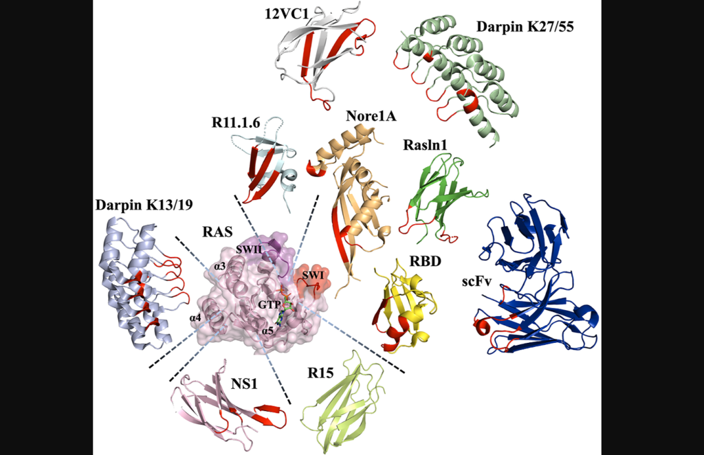
Although Ras has been considered difficult to target, recent advancements have identified potential binding pockets that can be addressed by small molecules, peptidomimetics and proteins. Inhibitors designed to covalently bind to the Ras G12C mutant have shown promise, leading to FDA-approved drugs for specific lung cancers. Additionally, protein-based inhibitors that target Ras and its interactions with effectors, regulatory proteins and guanine nucleotide exchange factors offer alternative strategies for therapeutic intervention. These developments have challenged the notion that Ras is “undruggable” and highlight the potential for effective treatments against various cancer types.

On July 1, 2023, researchers Atilio Tomazini and Julia M. Shifman from The Hebrew University of Jerusalem published a new review paper in Oncotarget, entitled, “Targeting Ras with protein engineering.” The authors provide an overview of the challenges associated with targeting Ras proteins with small molecules and discuss how protein engineering has emerged as a promising method to overcome these challenges.

“While the development of small-molecule Ras inhibitors has been reviewed elsewhere [40], we focus our review on protein-based Ras inhibitors, describing the methods for their engineering, various scaffolds used for inhibitor design, and prospects for delivery of the designed Ras inhibitors into the cellular cytoplasm, where Ras is located.”

Protein Engineering

Protein scaffolds offer alternative approaches to small molecule drugs for engineering protein-based inhibitors. Unlike small molecules, protein domains can bind to targets through large surface areas, providing high affinity and specificity. Antibodies, natural protein effectors and novel binding domains are commonly used as protein scaffolds. Antibodies can be engineered into smaller versions to overcome limitations, while natural effectors can be modified to enhance binding affinity. Novel binding domains, unrelated to the target protein, possess structural robustness and can be evolved to exhibit strong binding. All three classes of protein scaffolds have been utilized to engineer Ras binders and explore strategies to inhibit Ras oncogenesis.

“Interestingly, all classes of protein scaffolds, including antibodies, natural effectors, and novel binding domains, have been utilized for engineering of Ras binders, allowing scientists to target various sites on the Ras surface and to explore different strategies for inhibiting Ras oncogenesis […].”

Methods for engineering protein inhibitors can be categorized into experimental directed evolution and computational design, or a combination of both. Experimental techniques involve display technologies such as phage display, yeast surface display, ribosome display, and mRNA display. These methods allow for the construction of combinatorial libraries of protein mutants, which are then screened using the target protein as a selection “bait.” The selected binders are sequenced to identify high-affinity mutants. Negative selection steps can be incorporated to enhance specificity by eliminating binders to unwanted targets. The number of mutants that can be assayed depends on the display technology used, with each approach having its limitations.

In addition to experimental approaches, computational methods have been proposed for protein binder design. Computational design enables rational targeting of specific binding epitopes on the target protein. However, computationally designed binders often have weak initial binding affinities and require affinity maturation through experimental techniques. Computational methods have been successful in designing focused libraries for yeast surface display experiments, where small libraries of protein mutants are designed based on computational predictions. This approach narrows down the choices to the most promising mutants, facilitating directed evolution experiments. By combining computational and experimental approaches, protein inhibitors with superior affinity and specificity have been developed.

“We have summarized all the described engineered Ras protein-based binders and their properties in Table 1.”

The Future of Intracellular Transport for Ras Inhibitors

Efficient delivery of molecules that bind to intracellular Ras proteins is essential for suppressing pro-cancer pathways and promoting anti-cancer activities. To overcome the challenge of crossing the cell membrane, different strategies have emerged. One approach involves utilizing short cell-penetrating peptides (CPPs) that can be fused to the desired protein, allowing entry into cells through direct translocation or endocytosis. However, improving the release of cargo proteins from endosomes remains a hurdle. Supercharging proteins with positively charged surfaces or leveraging bacterial toxins with intrinsic delivery mechanisms are alternative methods for intracellular protein delivery. Additionally, coupling cargo proteins to nanoparticles or employing mRNA delivery systems have shown promise, although they have their own limitations.

These protein delivery techniques have been explored for targeting Ras inhibitors. For instance, a human IgG1 antibody was engineered to selectively bind to Ras-GTP, inhibiting downstream signaling. Fusion of Ras binding domains to CPPs demonstrated competitive inhibition of Ras/effector interactions. Furthermore, optimized bacterial secretion systems and lipid nanoparticle-encapsulated mRNA platforms have been employed for efficient intracellular delivery of Ras-binding molecules. These advancements open up possibilities for targeted cancer therapies and disease treatments by enabling effective delivery of Ras binders to their intracellular target, thus influencing cancer-related signaling pathways.

Conclusions

In summary, targeting Ras proteins, despite their historically challenging nature, has seen significant progress in recent years. Small molecules, peptidomimetics and protein-based inhibitors have emerged as potential strategies for inhibiting Ras oncogenesis. Protein engineering, utilizing various protein scaffolds such as antibodies, natural effectors and novel binding domains, offers alternative approaches to traditional small molecule drugs.

Experimental directed evolution and computational design, alone or in combination, have facilitated the development of high-affinity and specific protein inhibitors. Furthermore, the efficient intracellular delivery methods described above hold promise for targeted cancer therapies by effectively delivering Ras binders to their intracellular targets. These advancements challenge the perception of Ras as “undruggable” and provide hope for the development of effective treatments for various cancer types.

“These strategies should be utilized in future to examine the beneficial activity of Ras-binders and inhibitors and should further facilitate the development of protein-based Ras therapeutics.”

Click here to read the full review in Oncotarget.

ONCOTARGET VIDEOS: YouTube | LabTube | Oncotarget.com

Oncotarget is an open-access, peer-reviewed journal that has published primarily oncology-focused research papers since 2010. These papers are available to readers (at no cost and free of subscription barriers) in a continuous publishing format at Oncotarget.com. Oncotarget is indexed/archived on MEDLINE / PMC / PubMed.

Click here to subscribe to Oncotarget publication updates.

For media inquiries, please contact media@impactjournals.com.

Novel GEM Model Unveils PLK1’s Role in Tumorigenesis

In a new editorial, researchers discuss a study using their team’s new genetically engineered mouse (GEM) model to assess PLK1 as a driver of oncogenic transformation.

Figure 1: The role of PLK1 in tumorigenesis and cancer heterogeneity. GEM Model

On the bright side, polo-like kinase 1 (PLK1) is considered a master regulator of the ever-important cell cycle. On the dark side, PLK1 expression (at both the mRNA and protein level) has shown to be upregulated in tumor cells, suggesting that PLK1 may also contribute to tumorigenesis. Despite this direct association, researchers studying the role of PLK1 in cancer have encountered a problem: a lack of appropriate animal models for experimentation.

“Even though studies have suggested that PLK1 contributes to tumorigenesis, the ability of PLK1 to drive oncogenic transformation on its own in vivo was still questionable due to a lack of sophisticated animal models for experimentation [18, 19].”

This problem may have been solved in 2021. In a new editorial paper, researchers Lilia Gheghiani and Zheng Fu from Virginia Commonwealth University discuss a recent study using their team’s new genetically engineered mouse (GEM) model to assess the ability of PLK1 to be a sole driver of oncogenic transformation in vivo. Their editorial was published in Oncotarget’s Volume 14 on June 27, 2023, and entitled, “The dark side of PLK1: Implications for cancer and genomic instability.”

PLK1 in Tumorigenesis

“To address this important scientific question, we generated a new genetically engineered mouse (GEM) model using the CAGGS (cytomegalovirus (CMV) early enhancer/chicken β-actin) promoter to drive exogenous PLK1 expression, allowing its ubiquitous and robust gene expression in transgenic mice [20].”

In an effort to determine if PLK1 overexpression causes tumors, the researchers created a new GEM mouse model that expresses high levels of PLK1. These high levels caused various types of spontaneous tumors. The increased PLK1 levels caused defects in cell division and resulted in abnormal numbers of centrosomes and compromised cell cycle checkpoints. This allowed for the accumulation of chromosomal instability, leading to abnormal numbers of chromosomes and tumor formation. In human cancers, higher PLK1 expression was associated with an increase in genome-wide copy number alterations. Their study provides evidence that abnormal PLK1 expression can trigger chromosomal instability and tumor formation, suggesting potential therapeutic opportunities for cancers with chromosomal instability.

“In summary, this study provides a novel GEM model that recapitulates the increased PLK1 expression observed in many human cancers and demonstrates that PLK1 overexpression drives spontaneous tumor formation in multiples organs in mouse, revealing the dark side of PLK1 as a potent proto-oncogene.”

Conclusions

In conclusion, the limitations of previous studies on PLK1 and its role in cancer have been partially addressed by the development of the new GEM model created by these researchers. This model allowed the team to examine PLK1’s ability to drive oncogenic transformation in vivo. Their study demonstrates that overexpression of PLK1 leads to the formation of spontaneous tumors in multiple organs, highlighting the dark side of PLK1 as a potent proto-oncogene. The findings of this study provide valuable insights into the role of PLK1 in tumorigenesis and suggest potential therapeutic opportunities for cancers associated with chromosomal instability. This breakthrough in animal models opens up new avenues for further research in understanding the mechanisms underlying PLK1-related tumorigenesis and developing targeted therapies to combat cancer.

“Alternative therapeutic strategies, such as co-delivery systems using nanoparticles or combination therapies, are under development in order to enhance the efficacy of PLK1 inhibition [2528]. With expanding discoveries of PLK1 function and mechanisms of action, we hope that PLK1-targeted therapies will soon join the frontlines in the fight against cancer.”

Click here to read the full editorial paper in Oncotarget.

ONCOTARGET VIDEOS: YouTube | LabTube | Oncotarget.com

Oncotarget is an open-access, peer-reviewed journal that has published primarily oncology-focused research papers since 2010. These papers are available to readers (at no cost and free of subscription barriers) in a continuous publishing format at Oncotarget.com. Oncotarget is indexed/archived on MEDLINE / PMC / PubMed.

Click here to subscribe to Oncotarget publication updates.

For media inquiries, please contact media@impactjournals.com.

New Study Reveals Genetic Risk Factors for Cancer in Saudi Arabia

In a new study, researchers found that 38.4% of a cohort in Saudi Arabia carried pathogenic variants linked to hereditary cancer risk.

New Study Reveals Genetic Risk Factors for Cancer in Saudi Arabia

Familial cancer is a fearsome reality for millions of people worldwide. While some cases of familial cancer syndrome (FCS) may be influenced by shared environmental or lifestyle factors within a family, others are solely due to genetic mutations passed down through generations. This problem is especially prevalent in Saudi Arabia—where rates of familial cancer are among the highest in the world.

“Cancer increased in the Kingdom of Saudi Arabia by 136% between 1999 and 2015 [4].”

Approximately 20% of all Saudi Arabian cancer patients have a family history of cancer. This population is likely to carry mutant alleles, presenting an opportunity for further exploration and research. By studying these individuals and their genetic profiles, scientists and healthcare professionals can gain valuable insights into the genetic factors contributing to familial cancer in the Saudi Arabian population. This knowledge can help improve risk assessment, develop targeted prevention strategies, and potentially lead to more effective treatments for familial cancer cases. 

In a new study, researchers Musa AlHarbi, Nahla Ali Mobark, Wael Abdel Rahman AlJabarat, Hadeel ElBardis, Ebtehal AlSolme, Abdullah Bany Hamdan, Ali H. AlFakeeh, Fatimah AlMushawah, Fawz AlHarthi, Abdullah A. AlSharm, Ali Abdullah O. Balbaid, Naji AlJohani, Alicia Y. Zhou, Heather A. Robinson, Saleh A. Alqahtani, and Malak Abedalthagafi from King Fahad Medical City, Color Health Inc., University of Manchester, Johns Hopkins University, King Faisal Specialist Hospital and Research Center, and Emory University Hospital conducted a next-generation sequencing (NGS) assessment for hereditary cancer risk in a Saudi Arabian population. Their research paper was published in Oncotarget on June 12, 2023, entitled, “Investigating the prevalence of pathogenic variants in Saudi Arabian patients with familial cancer using a multigene next generation sequencing panel.”

The Study

The researchers used a 30-gene, targeted NGS panel to screen 310 subjects, including 57 non-cancer patients, 110 index patients with cancer and 143 of their relatives, 16 of whom also had cancer. (“Index patients” refers to individuals who are the first in a family to be diagnosed with a particular disease or condition of interest.) The NGS panel covered genes related to breast, ovarian, colorectal, endometrial, gastric, pancreatic, prostate, thyroid, renal, and skin cancers, as well as familiar adenomatous polyposis (FAP) and Lynch syndrome.

“This kit has been previously trialed as a means of capturing potential PVs [pathogenic variants] at a population level in Nigeria and the Caribbean, and in identifying rare variants in cancer patients who have tested negative for common cancer variants [3538].”

The results showed that 119 subjects (38.4% of the cohort) carried pathogenic or likely pathogenic variants (PVs) affecting genes associated with hereditary cancer risk. (TP53, ATM, CHEK2, CDH1, CDKN2A, BRCA1, BRCA2, PALB2, BRIP1, RAD51D, APC, MLH1, MSH2, MSH6, PMS2, PTEN, NBN/NBS1, and MUTYH were identified as genes with pathogenic or likely pathogenic variants.) Among 126 patients and relatives with a history of cancer, 49 subjects (38.9%) carried pathogenic or likely pathogenic variants. Two specific variants (APC c.3920T>A and TP53 c.868C>T) were significantly associated with the occurrence of colorectal cancer/Lynch syndrome and multiple colon polyposis. Diverse variants in BRCA2, many of which were previously unreported as pathogenic, were found at a higher frequency in individuals with a history of cancer compared to the general patient population. Overall, these subjects had more genetic variants associated with familial cancers compared to other populations.

Conclusion

“In conclusion, this study is one of the first to report the prevalence of inherited cancer genetic variants in a cohort from the Arab world. Our study gives critical first insights into the genetic variants associated with overall cancer risk in this specific population, and specific forms including CRC/Lynch syndrome and breast cancer.”

The researchers concluded that their study was the first to use a comprehensive NGS panel for FCS risk assessment in Saudi Arabia and that it provided valuable insights into the genetic landscape of cancer in this population. They also acknowledged some limitations of their study, such as the small sample size, the lack of clinical data for some subjects and the possibility of false negatives due to technical or analytical issues. Overall, this study highlighted the importance of genetic testing and counseling for FCS in Saudi Arabia, where consanguineous marriages are common and may increase the risk of inheriting cancer-associated alleles from both parents. These findings also suggested that knowing the genetic profile of patients and their families could help tailor preventive strategies and treatments according to their specific risks.

“Whilst a larger population level study is still needed, we demonstrate that multigene NGS panel testing may serve as non-invasive diagnostic and cost-effective tool to predict familial cancer risk at the pre-clinical stage, allowing targeted screening and enabling early intervention.”

Click here to read the full research paper in Oncotarget.

ONCOTARGET VIDEOS: YouTube | LabTube | Oncotarget.com

Oncotarget is an open-access, peer-reviewed journal that has published primarily oncology-focused research papers since 2010. These papers are available to readers (at no cost and free of subscription barriers) in a continuous publishing format at Oncotarget.com. Oncotarget is indexed/archived on MEDLINE / PMC / PubMed.

Click here to subscribe to Oncotarget publication updates.

For media inquiries, please contact media@impactjournals.com.

Can the Creatine Shuttle be Targeted to Fight Colorectal Cancer?

In a new study, researchers investigated the creatine shuttle pathway as a potential therapeutic target in colorectal cancer cells.

Can the Creatine Shuttle be Targeted to Fight Colorectal Cancer?

Listen to an audio version of this article

Since the 1992 Barcelona Olympics, creatine supplementation has increased in popularity and grown to widespread use among the mainstream public. Creatine is a naturally occurring compound, primarily stored in skeletal muscle and involved in energy production for high-intensity activities—enhancing performance and supporting muscle growth and strength. The process by which creatine is transported into the muscles and utilized for energy production is referred to as the creatine shuttle. While it is a useful mechanism for healthy muscles, the creatine shuttle has also been implicated in cancer.

“The creatine shuttle is highlighted in cancer as a source of energy for cancer cells that display aggressive proliferation, and aberrant creatine kinase (CK) levels are known to be associated with many malignancies and mitotic control [7].”

In a new study, researchers Mayu Kita, Rina Fujiwara-Tani, Shingo Kishi, Shiori Mori, Hitoshi Ohmori, Chie Nakashima, Kei Goto, Takamitsu Sasaki, Kiyomu Fujii, Isao Kawahara, Ujjal Kumar Bhawal, Yi Luo, and Hiroki Kuniyasu from Nara Medical University, Saveetha University and Nantong University hypothesized that the creatine shuttle is involved in energy metabolism and other adenosine triphosphate (ATP) supply in cancer cells. On May 19, 2023, their new research paper was published in Oncotarget’s Volume 14, entitled, “Role of creatine shuttle in colorectal cancer cells.”

“In the current study, the role of the creatine shuttle in CRC [colorectal cancer] was analyzed along with its potential as a therapeutic target.”

The Creatine Shuttle in Colorectal Cancer

Despite advancements in treatment options for colorectal cancer (CRC), incidence and mortality rates remain high. The metabolism of CRC cells is distinctly different from that of normal cells, and understanding these metabolic alterations is crucial for devising new targeted therapies. The creatine shuttle system plays a pivotal role in cellular energy metabolism, particularly in high-energy demanding tissues such as muscle and brain. However, its involvement in CRC cells has remained largely unexplored until now.

​​Creatine kinase, also known as CK or creatine phosphokinase, is an enzyme that catalyzes the transfer of a phosphate group from creatine phosphate to adenosine diphosphate (ADP), thereby regenerating adenosine triphosphate (ATP), which is the primary energy source for cells. CK exists in different forms or isoenzymes. In this study, the researchers investigated the expression and role of creatine kinase B (CKB) and mitochondrial creatine kinase (MTCK) in CRC tissues. They also explored the inhibitory effect of dinitrofluorobenzene (DNFB) on CKB and MTCK activity and its impact on CRC cell growth, stemness, mitochondrial function, energy metabolism, and cancer metastasis.

Inhibition of the Creatine Shuttle

The team used tissue arrays to examine CKB and MTCK expression in CRC tissues. Both proteins were highly expressed in high-grade tumors and cases with distant metastasis. Liver metastases showed higher expression compared to primary tumors, suggesting a role in CRC progression and metastasis.

DNFB, an inhibitor of CK activity, reduced CK activity and inhibited cell growth in CT26 and HT29 CRC cell lines. HT29 cells, with higher CKB and MTCK levels, were less sensitive to DNFB than CT26 cells. DNFB treatment decreased cell number, stem cell marker expression and impaired sphere formation in CT26 and HT29 cells. Knockdown of CKB or MTCK showed similar effects, indicating specificity to CK inhibition. DNFB also inhibited mitochondrial function and energy metabolism, decreasing mitochondrial membrane potential, increasing ROS production, and reducing OCR and ATP production in both cell lines.

In a mouse model of peritoneal dissemination, pretreatment with DNFB reduced tumor growth. Excised tumors from DNFB-treated mice showed decreased proliferation and stem cell marker expression, as well as reduced phosphorylation levels of tumor-promoting signaling molecules (EGFR, AKT, and ERK1/2).

Summary & Conclusion

“In this study, we showed that inhibition of the creatine shuttle by blocking CKB and MTCK activity suppressed the growth, stemness, and metastasis of cancer. It was suggested that the cause of this is related to inhibition of both mitochondrial energy metabolism and the phosphorylation signaling system.”

This research study provides valuable insights into the role of CKB and MTCK in CRC and highlights the therapeutic potential of inhibiting the creatine shuttle in CRC treatment. Inhibition of CKB and MTCK activity by DNFB impaired CRC cell growth, stemness, mitochondrial function, energy metabolism, and cancer metastasis. These findings suggest that targeting the creatine shuttle pathway may represent a promising therapeutic strategy for CRC patients. Further studies are warranted to validate these findings and explore the potential of targeting the creatine shuttle in clinical settings.

“Our data suggest that the antitumor effect of creatine shuttle inhibition can be attributed to the inhibition of mitochondrial energy production as well as the inhibition of multiple phosphorylation signals through inhibition of the ATP supply. Therefore, it is necessary to develop a new CK inhibitor to induce these two effects in vivo.”

Click here to read the full research paper in Oncotarget.

ONCOTARGET VIDEOS: YouTube | LabTube | Oncotarget.com

Oncotarget is an open-access, peer-reviewed journal that has published primarily oncology-focused research papers since 2010. These papers are available to readers (at no cost and free of subscription barriers) in a continuous publishing format at Oncotarget.com. Oncotarget is indexed/archived on MEDLINE / PMC / PubMed.

Click here to subscribe to Oncotarget publication updates.

For media inquiries, please contact media@impactjournals.com.

Reaching the Brain Through the Groin: A Novel Approach to Brain Cancer

In a new editorial, researchers discuss opening the blood-brain barrier and a promising new strategy for the treatment of brain cancer.

Figure 1: A transfemoral path to BBB opening.

Listen to an audio version of this article

Just a small number of molecules, including alcohol and caffeine, are able to cross the blood-brain barrier (BBB). The BBB is a highly selective semipermeable membrane that separates circulating blood from extracellular fluid in the brain. It plays a critical role in protecting the brain from harmful substances in the blood while also maintaining a stable and consistent environment for neuronal function. Without the BBB, humans would be at the mercy of any harmful toxin, pathogen and unwanted substance that could cross from the bloodstream into the brain.

This protective function also makes it difficult to deliver therapeutic agents to the brain, as the majority of drugs and other molecules are unable to cross the BBB. This is particularly problematic for the treatment of brain-localized diseases, including brain cancers and neurological disorders, which require high concentrations of drugs to effectively target sites in the brain. In a new editorial paper, researchers Thomas C. Chen, Weijun Wang and Axel H. Schönthal from the University of Southern California‘s Keck School of Medicine discuss a series of preclinical studies that introduced the novel concept of intraarterial (IA) injection of NEO100—a promising strategy aimed at temporarily and safely opening the BBB up for therapeutic treatment. Their editorial was published in Oncotarget’s Volume 14 on May 4, 2023, entitled, “From the groin to the brain: a transfemoral path to blood-brain barrier opening.”

“It is believed that procedures to open the BBB in a controlled and safe fashion might provide tremendous advantages by allowing optimal brain entry of any and all circulating therapeutics.”

Opening the BBB

The authors first describe previously used methods of opening the BBB for therapeutic intervention, including intracarotid injection of hyperosmolar mannitol and MRI-guided pFUS with intravascular microbubbles. Unfortunately, these methods have yielded issues with safety and efficacy. Fortunately, Chen, Wang, Schönthal, and their co-authors came up with a new idea for opening the BBB safely. 

In a 2021 study, the researchers discovered that NEO100 enables the delivery of BBB-impermeable therapeutics to the brain. NEO100 is a type of perillyl alcohol—a natural chemical found in citrus fruit peels—that has been studied for its potential to treat cancer. Wang et al. aimed to see if injecting NEO100 into an artery would open the BBB safely and temporarily. This could help other drugs that are normally unable to pass through the BBB, such as methotrexate and therapeutic antibodies, to enter the brain. Previously, NEO100 had been administered through the nose to treat cancer, but this study focused on its ability to open the BBB.

The researchers injected NEO100 into the left ventricle of the heart and then injected a dye called Evans blue into the mice’s veins. Normally, this dye cannot penetrate the brain, but when the BBB is weakened or opened up, it can get through and turn the brain blue. And that’s exactly what happened—the mice’s brains turned blue after the injections. Interestingly, when they tried using another substance called mannitol, it did not have the same effect on the BBB. The team performed additional studies and found that NEO100 seemed to affect the connections between cells in the barrier.

In further experiments, the researchers used methotrexate and special markers that usually do not enter the brain. They gave these drugs and markers to mice and found that NEO100 made it easier for the drugs and markers to enter the brain. This effect lasted between two and four hours before the BBB reverted to normal functioning. The researchers also tested administering NEO100 by injecting it into the mouse’s veins, but this was not effective. 

The main question the researchers wanted to answer was if opening the BBB using IA NEO100 could help treat brain tumors. To answer this question, they conducted experiments using mice that had tumor cells implanted in their brains. In one study, they used breast cancer cells that were engineered to have the protein HER2 and treated them with trastuzumab. In another study, they used models of brain cancer called melanoma and glioblastoma and treated them with drugs that help the immune system fight cancer. These studies have found a way to improve drug delivery for CNS diseases, but there are limitations that need further investigation.

Transfemoral IA catheterization

As noted in this editorial, the preclinical models above used one injection of NEO100 with a therapeutic agent, but it’s unclear if this will work as well in humans. Tumors in humans are more complex than in rodents, so multiple interventions might be needed. It is also important to determine the best way to perform the injection(s) in humans. The researchers suggest using a catheter inserted through the femoral artery near the groin and guided by fluoroscopy to safely inject NEO100 into the cranial arteries.

“Transfemoral IA catherization (Figure 1) is a low-risk procedure that is routinely performed by endovascular neurosurgeons in the context of cerebral angiograms, aneurysm coiling, tumor embolization, and thrombectomies [18]. It is considered ‘the gold standard technique for catheter-based neuro-interventions’ [19]. However, it has never been used as a means to access tumor-feeding cranial arteries for purposes of BBB opening.”

Transfemoral IA catheterization is a medical procedure that involves inserting a catheter through a blood vessel in the leg and guiding it to the brain to perform various treatments. It is a safe and common technique, already used by doctors who specialize in treating brain conditions. However, it has never been used to open the BBB in order to access the blood vessels. Using NEO100 with this procedure could be a new and innovative way to treat aggressive brain tumors. If necessary, the procedure could even be repeated multiple times due to its safe and simplistic nature. The researchers believe that using this new method to open the BBB could be just as successful in treating brain tumors as current treatments are for tumors in other parts of the body. This could potentially lead to better outcomes for patients with brain tumors, such as improved survival rates and fewer side effects.

Conclusions

The blood-brain barrier (BBB) is a protective barrier that prevents harmful substances from entering the brain. However, this barrier also makes it difficult to deliver therapeutic agents to the brain. In a new study, researchers have proposed a novel method of intraarterial injection of NEO100 to temporarily and safely open the BBB. This method has been shown to enable the delivery of BBB-impermeable therapeutics to the brain. The authors of this editorial have suggested using transfemoral IA catheterization to perform this intervention. The method requires further investigation and development.

“The authors envision that clinical implementation of this new BBB-opening method might achieve a similarly high rate of success in the treatment of brain-localized malignancies as do current treatments for peripherally distributed tumors; as a result, reduced morbidity and increased patient survival is expected.”

Click here to read the full editorial in Oncotarget.

ONCOTARGET VIDEOS: YouTube | LabTube | Oncotarget.com

Oncotarget is an open-access, peer-reviewed journal that has published primarily oncology-focused research papers since 2010. These papers are available to readers (at no cost and free of subscription barriers) in a continuous publishing format at Oncotarget.com. Oncotarget is indexed/archived on MEDLINE / PMC / PubMed.

Click here to subscribe to Oncotarget publication updates.

For media inquiries, please contact media@impactjournals.com.

Defining the Complexity of EGFR Endocytosis in Cancer

In a new editorial perspective, researchers delve into the complex mechanisms underlying EGFR endocytosis and its potential as a therapeutic target.

Figure 1: SNX3 protein downregulation in breast tumors.

The Trending With Impact series highlights Oncotarget publications attracting higher visibility among readers around the world online, in the news and on social media—beyond normal readership levels. Look for future science news about the latest trending publications here, and at Oncotarget.com.

Listen to an audio version of this article

EGFR (epidermal growth factor receptor) is a crucial protein that plays a significant role in various biological processes such as cell growth, proliferation, differentiation, and survival. Dysregulation of EGFR signaling has been implicated in the development and progression of numerous human cancers, including lung, breast and colon cancer. Therefore, EGFR has emerged as an attractive target for cancer therapy, and several drugs that target EGFR are in clinical use or under investigation.

In recent years, endocytosis, the process by which cells internalize molecules and transport them into intracellular compartments, has emerged as a critical modulator of EGFR signaling. Endocytosis of EGFR not only regulates the duration and intensity of EGFR signaling but also modulates the signaling output. Dysregulation of EGFR endocytosis has been implicated in the development of drug resistance to EGFR-targeted therapies, highlighting the importance of understanding the mechanisms that regulate EGFR endocytosis.

In a new editorial perspective, researchers Aysegul Sapmaz and Ayse Elif Erson-Bensan from Middle East Technical University provide an overview of the recent advances in our understanding of EGFR endocytosis and its role in EGFR signaling and cancer. The authors highlight the importance of the dynamic interplay between EGFR endocytosis and downstream signaling pathways and discuss how aberrant EGFR endocytosis contributes to drug resistance to EGFR-targeted therapies. On April 10, 2023, their editorial perspective was published in Oncotarget’s Volume 14, entitled, “EGFR endocytosis: more than meets the eye.”

“Here we review the role of the EGF-SNX3-EGFR axis in breast cancers with an extended discussion on deregulated EGFR endocytosis in cancer.”

EGFR Endocytosis

In a recent 2022 study, Sapmaz, Erson-Bensan and their team made significant contributions to understanding the role of deregulated endocytosis in cancer by describing the tumor suppressor role of Sorting Nexin 3 (SNX3) in triple-negative breast cancers (TNBCs). SNX3 is a protein-coding gene belonging to a family of proteins called sorting nexins, which are involved in sorting and trafficking of cellular membrane proteins and lipids. At the conclusion of their study, the researchers found that SNX3 is a critical player in TNBCs through the EGF-SNX3-EGFR axis.

“SNX3, an endosomal trafficking protein, is an emerging tumor suppressor in breast cancers as a target of the EGFactivated EGFR pathways and a modulator of EGFR protein levels.”

In the current editorial perspective by Sapmaz and Erson-Bensan, they discuss overexpression of EGFR and its activating mutations linked to various cancer phenotypes, including stemness, metastasis and drug resistance. Endocytosis and the internalization of EGFR play a crucial role in regulating its activity, which is dependent on post-translational modifications and regulated by various proteins, including ubiquitin. Deregulation of these players in endocytic processes has significant implications for EGFR activity in cancers. The ubiquitination status of EGFR and other proteins in the endocytic pathway is functionally essential and is balanced by E3 ubiquitin ligases and deubiquitinating enzymes (DUBs). Targeting these enzymes to alter ubiquitination dynamics could offer future perspectives in manipulating EGFR endocytosis and signaling in cancers.

The researchers discuss the non-canonical functions of endocytosis and endocytosis-related proteins, such as their involvement in nucleocytoplasmic shuttling and transcriptional activity. Several endocytic proteins have been found to interact with nuclear proteins and modulate gene transcription. Examples include EPS15, EPN1 and RNF11, which can shuttle between the cytoplasm and nucleus and have been shown to positively regulate transcription. Other endocytic proteins and adaptors have also been found to shuttle between the cytoplasm and nucleus with functions in endocytosis and gene expression. The authors emphasize the need for a better understanding of both the canonical and non-canonical functions of endocytic processes for normal physiology and diseases, including cancer and other pathologies.

“A better understanding of these backstage mechanisms will allow a more comprehensive understanding of receptor fate and activity.”

Conclusions

In conclusion, EGFR and its regulation through endocytosis have emerged as critical players in cancer development and progression. Dysregulation of EGFR endocytosis has been implicated in drug resistance to EGFR-targeted therapies, highlighting the importance of understanding the mechanisms that regulate this process. Recent advances in our understanding of EGFR endocytosis and its role in cancer have revealed the critical interplay between EGFR signaling and downstream pathways. The research by Sapmaz and Erson-Bensan sheds light on the tumor suppressor role of SNX3 in TNBCs and highlights the need for a better understanding of the canonical and non-canonical functions of endocytic processes in normal physiology and diseases, including cancer. Future research focusing on manipulating EGFR endocytosis and signaling could offer new perspectives on cancer therapy.

“Finally, before we can consider key endocytosis regulators as therapeutic targets, these candidate proteins must also be evaluated within the context of potential feedback mechanisms to modulate the biosynthesis and repopulation of receptors in cancer cells.”

Click here to read the full editorial perspective in Oncotarget.

ONCOTARGET VIDEOS: YouTube | LabTube | Oncotarget.com

Oncotarget is an open-access, peer-reviewed journal that has published primarily oncology-focused research papers since 2010. These papers are available to readers (at no cost and free of subscription barriers) in a continuous publishing format at Oncotarget.com. Oncotarget is indexed/archived on MEDLINE / PMC / PubMed.

Click here to subscribe to Oncotarget publication updates.

For media inquiries, please contact media@impactjournals.com.

The Evolution of Metastatic Cancer: Mechanisms and Drivers

In a new editorial, researchers explore genomic evolution in metastatic cancer, how therapy can drive it and the implications for developing new treatments. 

The Evolution of Metastatic Cancer: Mechanisms and Drivers

The Trending With Impact series highlights Oncotarget publications attracting higher visibility among readers around the world online, in the news and on social media—beyond normal readership levels. Look for future science news about the latest trending publications here, and at Oncotarget.com.

Listen to an audio version of this article

There are several theories that attempt to explain the genesis of cancer. One prominent theory is the genetic theory—proposing that cancer may arise from the accumulation of genetic mutations that alter the normal functioning of cells. These mutations can drive the formation of tumors, which can then spread to other parts of the body in a process known as metastasis. Metastatic cancer is often difficult to treat because it has evolved to become resistant to standard therapies. 

“It is generally accepted that development of cancer is a slow process, likely spanning decades during which the developing neoplastic cells sequentially acquire genomic alterations that will eventually give rise to the primary tumor [1].”

In a new editorial, researchers Ditte S. Christensen and Nicolai J. Birkbak from Aarhus University discuss mechanisms of genomic evolution in metastatic cancer, how therapy can drive it and the implications for developing new treatments. Their editorial paper was published in Oncotarget on March 21, 2023, entitled, “Therapy drives genomic evolution in metastatic cancer.”

Therapy Can Drive Metastatic Cancer

Cancer cells are master adaptors and have a remarkable ability to evolve, especially in response to therapy. When cancer cells are exposed to chemotherapy, radiation or targeted therapies, they can develop resistance to these treatments by acquiring new genetic mutations. This can occur through a variety of mechanisms, including mutations in the genes that regulate cell division and DNA repair, as well as the acquisition of new genes that confer resistance to specific drugs.

In this editorial, the authors discuss how this process of genomic evolution can lead to the development of metastatic cancer. As cancer cells acquire new mutations that allow them to survive and grow in the presence of therapy, they may also acquire mutations that allow them to invade and colonize new tissues. This can lead to the development of new tumors in distant parts of the body, which are often more difficult to treat than the original tumor.

“How the ability to perform these multiple independent steps is acquired by cancer cells remains a mystery.”

Mechanisms of Metastatic Cancer

Understanding the mechanisms by which cancer cells evolve in response to therapy is essential for developing new treatments for metastatic cancer. The clonal bottleneck hypothesis and the gatekeeper mutation hypothesis are two different hypotheses that attempt to explain how cancer cells acquire the ability to metastasize and spread to distant parts of the body. The clonal bottleneck hypothesis proposes that metastatic cancer is the result of a single subclone of cancer cells from the primary tumor that successfully seeds new sites. According to this hypothesis, the cancer cells undergo a clonal bottleneck event where only a small number of cells from the primary tumor are able to survive and successfully colonize new tissues. This hypothesis suggests that the ability to metastasize is an inherent property of the subclone that successfully colonizes new sites.

On the other hand, the gatekeeper mutation hypothesis proposes that metastatic cancer is the result of a specific genetic mutation or mutations that act as gatekeepers, allowing cancer cells to metastasize and spread to new sites. According to this hypothesis, the ability to metastasize is acquired through the acquisition of one or more specific genetic mutations that allow cancer cells to bypass the normal checks and balances that prevent uncontrolled growth and invasion of surrounding tissues.

Exploring Gatekeeper Genomic Events

In a 2022 study, the authors of this editorial and their team explored the concept of gatekeeper genomic events by comparing primary and metastatic tumors on a large scale. Their large-scale analysis of more than 40,000 individual tumors from the AACR Genomics Evidence Neoplasia Information Exchange (GENIE) project found an increase in mutation burden and chromosomal instability in metastatic tumors, but no evidence of individual mutations driving the metastatic process itself. The concept of gatekeeper mutations remains a hypothesis, and further research is needed to explore this idea in more detail.

This study and others suggest that metastatic cancer dissemination involves a bottleneck event where a highly fit clone from a primary tumor successfully seeds distant sites. Strong selective pressure from anti-cancer therapy drives the acquisition of private driver mutations associated with therapy resistance in individual metastatic tumors. There is limited evidence for the existence of specific gatekeeper mutations. It is also possible that the primary driver of metastatic cancer is found outside the cancer cells.

“Indeed, it may be that a primary driver of metastatic cancer is to be found outside the cancer cells themselves, potentially through inflammation in the tumor-immune microenvironment or through interaction with a declining host immune system which may enable immune escape and sudden systemic dissemination by a highly proliferative primary tumor clone.”

Conclusions

In conclusion, the genetic theory of cancer proposes that cancer arises from genetic mutations that alter the normal functioning of cells, leading to the formation of tumors and metastasis. This editorial by Christensen and Birkbak highlights the process of genomic evolution in metastatic cancer and its implications for cancer treatment. Understanding the mechanisms by which cancer cells evolve in response to therapy is crucial for developing new treatments for metastatic cancer. Recent studies have shed light on the clonal origin of metastatic tumors and the role of selective pressure from anti-cancer therapy in the acquisition of private driver mutations associated with therapy resistance. While the concept of gatekeeper mutations remains a hypothesis, it is clear that the acquisition of aggressive cancer traits is the primary driver of metastatic potential. Further research is needed to explore this idea in more detail and develop effective therapies for metastatic cancer.

“It will be exciting to further explore these questions as more data becomes available on metastatic cancers, particularly with paired primary and metastatic tumor samples with sequential biopsies to facilitate the analysis of dynamic tumor evolution over time, rather than through static snapshots provided by samples obtained at a single time point.”

Click here to read the full editorial published in Oncotarget.

ONCOTARGET VIDEOS: YouTube | LabTube | Oncotarget.com

Oncotarget is an open-access, peer-reviewed journal that has published primarily oncology-focused research papers since 2010. These papers are available to readers (at no cost and free of subscription barriers) in a continuous publishing format at Oncotarget.com. Oncotarget is indexed/archived on MEDLINE / PMC / PubMed.

Click here to subscribe to Oncotarget publication updates.

For media inquiries, please contact media@impactjournals.com.

Targeting Pre-Leukemic Cells: New Hope for Preventing Childhood B-ALL

Researchers from Universidad Autónoma de Madrid published a new editorial in Oncotarget detailing a proof-of-principle experiment to prevent B-cell acute lymphoblastic leukemia (B-ALL).

Researchers from Universidad Autónoma de Madrid published a new editorial in Oncotarget detailing a proof-of-principle experiment to prevent B-cell acute lymphoblastic leukemia (B-ALL).

The Trending With Impact series highlights Oncotarget publications attracting higher visibility among readers around the world online, in the news, and on social media—beyond normal readership levels. Look for future science news about the latest trending publications here, and at Oncotarget.com.

Listen to an audio version of this article

Childhood leukemia is a devastating disease that affects thousands of children every year. Despite significant advancements in the field of pediatric oncology, childhood leukemia remains a major cause of morbidity and mortality in children, with B-cell acute lymphoblastic leukemia (B-ALL) being the most common form. 

Some cases of childhood B-ALL arise from congenital mutations that lead to a silent population of pre-leukemic cells. These cells at some point are triggered by a catalyst (possibly by delayed exposure to a common infection), acquire additional genetic mutations and ultimately develop into B-ALL. However, researchers have yet to fully understand how to target these pre-leukemic cells to prevent B-ALL.

In a new editorial paper, researchers César Cobaleda, Manuel Ramírez-Orellana, Carolina Vicente-Dueñas, Andreas Weiss, Kim E. Nichols, and Isidro Sánchez-García from Universidad Autónoma de Madrid discuss a novel method of targeting pre-leukemic cells in practice. On March 11, 2023, the team published their editorial in Oncotarget, entitled, “Proof-of-principle: targeted childhood leukemia prevention.” 

“[…] one would have to find a way to specifically target these preleukemic cells. Recently, a mouse model recapitulating the phenotype of a leukemia-predisposition syndrome has allowed us to carry out a proof-of-principle experiment to achieve this very goal.”

The Study

Pax5 is a protein that plays a critical role in the development of white blood cells that produce antibodies to fight infections, B cells. A Pax5 mutation or deficiency can lead to a disruption in the development of B cells and compromise the immune system’s ability to fight off infections. Pax5+/- refers to an individual or organism that has one functional copy of the Pax5 gene and one non-functional copy. This condition is also known as heterozygosity.

“Children carrying heterozygous mutations affecting the B-cell master regulator gene PAX5 are predisposed to develop B-ALL; similarly, 25% of heterozygous Pax5+/− mice develop leukemia, but only after experiencing an immune stress, such as exposure to infection [24].”

In their recent study, the researchers used Pax5+/− mice (a mouse model carrying a leukemia-predisposition syndrome) to evaluate whether in vivo treatment with ruxolitinib, a Jak1/2 inhibitor, administered early in life is capable of killing pre-leukemic cells and preventing the development of acute leukemia. They found that treatment with ruxolitinib led to the disappearance of B-cell progenitors in Pax5+/-, but not in wild-type (WT), mice. When both experimental Pax5+/- and control WT animals were fed with ruxolitinib-containing chow for 14 or 28 days and then exposed to common mouse pathogens, the animals treated with ruxolitinib for 28 days showed a significant 90% reduction in the incidence of B-ALL compared to untreated mice or animals treated only for 14 days. 

Ultra-deep sequencing studies of Pax5+/- mice showed that ruxolitinib acts by eliminating predisposed preleukemic B cells before the second “hit” (or catalyst) that leads to their descent into B-ALL. These findings suggest that an analogous approach could be used to prevent the development of B-ALL in children who carry germline mutations that predispose them to this disease.

“It is becoming increasingly clear that the existence of latent pretumoral cells is common to many types of both hematologic and solid cancers [7]; therefore, the concept described here could be considered a proof-of-principle strategy for the development of similar prophylactic approaches to prevent the progression of other malignancies.”

Figure 1: Targeted prevention of progression to B-ALL.

Summary & Conclusion

Childhood leukemia, specifically B-cell acute lymphoblastic leukemia, remains a significant cause of morbidity and mortality in children, despite advancements in pediatric oncology. The pre-leukemic cells that predispose children to B-ALL are not fully understood, making it challenging to prevent the disease. However, the recent study by Cobaleda C, et al. offers new hope. The proof-of-principle experiment demonstrates that early treatment with ruxolitinib can eliminate pre-leukemic cells and significantly reduce the incidence of B-ALL in the Pax5+/- mouse model.

Their findings suggest that a similar approach could be used to prevent B-ALL in children with germline mutations that predispose them to this disease. The concept described in this study could also serve as a strategy for developing prophylactic approaches to prevent the progression of other malignancies that share the existence of latent pretumoral cells. While further research is necessary, this study offers new possibilities in preventing childhood leukemia and improving the outcomes for children at risk.

“Still, some aspects remain unclear. For example, why do the majority of genetically predisposed animals (and most genetically predisposed children) not develop leukemia and stay healthy? In addition, what are the mechanisms by which environmental factors such as infection promote the acquisition of secondary mutations leading to malignant progression of preleukemic cells? These and other important questions still need to be answered if we are to fully understand and avert the appearance of B-ALL.”

Click here to read the full editorial published in Oncotarget.

ONCOTARGET VIDEOS: YouTube | LabTube | Oncotarget.com

Oncotarget is an open-access, peer-reviewed journal that has published primarily oncology-focused research papers since 2010. These papers are available to readers (at no cost and free of subscription barriers) in a continuous publishing format at Oncotarget.com. Oncotarget is indexed/archived on MEDLINE / PMC / PubMed.

Click here to subscribe to Oncotarget publication updates.

For media inquiries, please contact media@impactjournals.com.

Unconventional Protein Secretion (UPS) in Cancer

Researchers from The Jikei University School of Medicine published a recent editorial perspective in Oncotarget, entitled, “Leakage? or Secretion? unconventional protein secretion in cancer.” 

unconventional protein secretion in cancer
3D rendering of structure/anatomy of human cell with organelles, nucleus, membrane, mitochondria, Golgi apparatus

The Trending With Impact series highlights Oncotarget publications attracting higher visibility among readers around the world online, in the news, and on social media—beyond normal readership levels. Look for future science news about the latest trending publications here, and at Oncotarget.com.

Listen to an audio version of this article

Conventional protein secretion is fairly well understood in this day and age. Proteins that are secreted through the classical, secretory vesicle-related pathway contain a signal peptide that guides them to the endoplasmic reticulum (ER), where they are further processed and transported to the Golgi apparatus. From there, the Golgi apparatus modifies, sorts and packages proteins for transport to their final destinations within the cell or for secretion outside of the cell. 

However, in recent years, it has become clear that not all secreted proteins follow this conventional pathway. Instead, some proteins are secreted through unconventional pathways that do not require a signal peptide or the involvement of the ER or Golgi apparatus. In a recent editorial perspective published in Oncotarget on February 20, 2023, “Leakage? or Secretion? unconventional protein secretion in cancer,” researchers Kohji Yamada and Kiyotsugu Yoshida from The Jikei University School of Medicine in Tokyo, Japan, discuss unconventional protein secretion (UPS) and its significance in the progression of cancer. 

“In contrast, the secretion of proteins that do not code for signal peptides may occur via two or more mechanisms, often collectively referred to as unconventional protein secretion (UPS).”

UPS in Cancer

The mechanism of action of UPS in cancer cells is still unclear, however, UPS may be carried out through multiple mechanisms. One such mechanism is the lysosome-mediated secretion pathway, in which proteins are secreted through lysosome organelles that break down and recycle cellular waste. Another mechanism is the exosome-mediated secretion pathway, in which proteins are packaged into small vesicles called exosomes and released from the cell.

In their editorial perspective, Yamada and Yoshida focus on the unconventional secretion of proteins in cancer cells. They note that these unconventional pathways have been implicated in the progression of various types of cancer, especially in liver cancers. The authors quickly began to discuss the question in the title of their paper: leakage or secretion? Since cancer cells are often surviving in harsh external environments, they tend to undergo damage to their cellular membranes. This makes it difficult to identify whether the proteins found in the extracellular environment have been leaked or secreted from the cell.

“Particularly, in liver cancer, cytosolic proteins are secreted actively, but not just leaked. In our experiments with liver cancer cell lines, we demonstrated that proliferating cells under normal culture conditions release importin α1 and PKCδ [2, 3].”

The researchers then delve into the different proteins secreted through UPS and their role in cancer. They note that cancer cells often secrete proteins that promote tumor growth and metastasis through these unconventional pathways. For example, the lysosome-mediated secretion of the protein cathepsin B has been shown to promote cancer cell invasion and metastasis. Exosome-mediated secretion of various proteins, including growth factors and cytokines, has been linked to tumor growth and immune evasion.

Conclusion

Overall, the editorial perspective by Yamada and Yoshida provides valuable insight into the significance of unconventional protein secretion in cancer. While further research is needed in order to understand UPS, this editorial perspective highlights its potential as a therapeutic target. The authors emphasize the importance of continuing research into this area of cancer biology.

“In conclusion, we and other researchers have accumulated a series of evidences on UPS and its biological significance in cancer. However, the mechanism of action of UPS in cancer cells remains unclear. How cytosolic proteins localized to the ER via E-Syt1 migrate into vesicles for secretion or whether their secretory mechanisms involve other organelles, including the Golgi apparatus, autophagosomes, or lysosomes, remains unclear. In the future, if the route of UPS that is centered on E-Syt1 is elucidated, this will presumably lead to not only a basic understanding of liver cancer but also to the development of diagnostic and therapeutic strategies.”

Click here to read the full editorial perspective published in Oncotarget

ONCOTARGET VIDEOS: YouTube | LabTube | Oncotarget.com

Oncotarget is an open-access, peer-reviewed journal that has published primarily oncology-focused research papers since 2010. These papers are available to readers (at no cost and free of subscription barriers) in a continuous publishing format at Oncotarget.com. Oncotarget is indexed/archived on MEDLINE / PMC / PubMed.

For media inquiries, please contact media@impactjournals.com.

The Role of Kras and Canonical Wnt Pathways in Biliary Tract Cancers

In a recent Oncotarget editorial, researchers discuss Kras and the canonical Wnt pathway in biliary tract cancers, and potential theraputic strategies using these targets.

Figure 1: The role of Kras and canonical Wnt pathways for tumorigenesis of extrahepatic biliary system.
Listen to an audio version of this article

The extrahepatic biliary system is a network of tubes and ducts that carry bile from the liver to the small intestine, where it helps digest fats. Biliary tract cancers, including gallbladder cancer and cholangiocarcinoma, are rare but aggressive cancers that arise from this system. Understanding the molecular mechanisms that drive these cancers is crucial for developing effective therapies.

“Despite advances in diagnosis and therapy, 5-year survival rate of biliary cancer is only 5% to 15% [7, 8].”

In a new editorial, researchers Munemasa Nagao, Akihisa Fukuda and Hiroshi Seno from Kyoto University Graduate School of Medicine discuss the latest research on the role of Kras and the canonical Wnt pathway in the development of biliary tract cancers. On January 26, 2023, their paper was published in Oncotarget’s Volume 14, entitled, “The role of Kras and canonical Wnt pathways for tumorigenesis of extrahepatic biliary system.”

Kras and The Canonical Wnt Pathway in Biliary Tract Cancers

Kras is a gene that plays a key role in regulating cell growth and division. Mutations in Kras are common in many types of cancer, including biliary tract cancers. The authors of this editorial note that recent studies have shown that Kras mutations are relatively frequent in biliary cancers. These mutations activate the Kras protein, leading to uncontrolled cell growth and division.

The canonical Wnt pathway is another molecular pathway that has been implicated in cancer development. The Wnt pathway helps regulate cell growth and division during embryonic development and in adult tissues. Abnormal activation of the Wnt pathway has been linked to several types of cancer, including colon cancer and liver cancer. Recent studies have shown that the canonical Wnt pathway is also activated in biliary tract cancers.

“However, the role of the KRAS and WNT pathways in biliary tumorigenesis remained unclear.”

A protein called beta-catenin, which is a key component of the Wnt pathway, is often overexpressed in biliary tract cancers. This leads to the activation of downstream target genes that promote cell growth and division. The researchers also discussed results from their 2022 study investigating the role of the Kras and canonical Wnt pathways in the tumorigenesis of the extrahepatic biliary system using a genetically engineered mouse (GEM) model.

“In summary, concurrent activation of the Kras and Wnt pathways in the extrahepatic biliary system induced ICPN and BilIN, which can progress to biliary cancer (Figure 1). This study provides the first novel GEM that recapitulates human ICPN and BilIN, establishing them precancerous lesions. This work shows how dysregulation of canonical cell growth pathways drives precursors to biliary cancers and identifies several molecular vulnerabilities as potential therapeutic targets in these precursors to prevent oncogenic progression.” 

Potential Therapeutic Strategies for Biliary Tract Cancers

The researchers go on to discuss the concurrent activation, or crosstalk, between Kras and these pathways in biliary tract cancers. They note that several studies have shown that Kras mutations can activate the Wnt pathway, leading to even more aggressive cancer growth. This suggests that targeting both pathways may be necessary for effective therapy.

Potential therapeutic strategies targeting Kras and Wnt pathways in biliary tract cancers were discussed in this editorial. Several drugs that target these pathways are currently in development, and some are already being tested in clinical trials. The authors suggest that combining these drugs with chemotherapy or other targeted therapies may be a promising approach for treating biliary tract cancers.

“To develop novel preventive and therapeutic approaches for extrahepatic biliary cancer, it is also important to clarify the role of other altered genes by using a GEM model and/or human samples.”

Conclusion

Overall, this editorial provides a comprehensive overview of the latest research on the molecular mechanisms underlying biliary tract cancers. Their discussion of the role of Kras and the canonical Wnt pathway highlights the importance of understanding these pathways for developing effective therapies. The potential for combination therapies targeting both Kras and the canonical Wnt pathway is particularly intriguing, and could offer new hope for patients with these aggressive cancers.

Withstanding, it is important to note that more research is needed before these ideas can be translated into clinical practice. The authors themselves acknowledge that the development of effective targeted therapies for biliary tract cancers is still in its early stages. However, with continued research and collaboration, it is possible that new treatments will emerge that can improve the prognosis for patients with these challenging cancers.

Click here to read the full editorial published in Oncotarget

ONCOTARGET VIDEOS: YouTube | LabTube | Oncotarget.com

Oncotarget is an open-access, peer-reviewed journal that has published primarily oncology-focused research papers since 2010. These papers are available to readers (at no cost and free of subscription barriers) in a continuous publishing format at Oncotarget.com. Oncotarget is indexed/archived on MEDLINE / PMC / PubMed.

For media inquiries, please contact media@impactjournals.com.