Mikhailblagosklonnyoncotarget.com features weekly articles describing new and trending research papers published by Oncotarget—an open access biomedical journal dedicated to the publishing of the latest oncology-focused, peer-reviewed research papers.
Authored by the Scientific Integrity Office at Oncotarget, the editorial comprehensively analyzes the journal’s evolving approach to scientific integrity. It addresses historical challenges in scholarly publishing and discusses the necessity of modern image forensics tools to meet the most rigorous standards of scientific integrity.
The Scientific Integrity Office describes how advances in digital technologies—particularly image forensics tools such as ImageTwin and analytical platforms like Argos—have transformed the ability to detect problematic data and analyze the quality of published research.
The editorial emphasizes that the lack of adequate image tools in the “pre-tools” era limited journals’ ability to detect image-related issues, underscoring the importance of recent technological advancements. It also highlights that Argos provides a good opportunity to obtain an objective picture across different journals in both the pre- and post-tools eras.
Looking forward, Oncotarget advocates indexes for broader adoption of independent analytical and AI-based tools in journal evaluation. In the public interest, it also encourages open discussion of how indexes select, deselect, and reevaluate journals.
“This article is a contribution to the special issue of Aging celebrating the life and work of Misha Blagosklonny (more formally, Mikhail Vladimirovich Blagosklonny), who died in October 2024.”
In this review, David Gems and Alexander Carver from University College London, together with Yuan Zhao from Queen Mary University of London, present a new theoretical model to explain how aging leads to the development of chronic diseases. Drawing on evolutionary theory and biological research, the authors propose that aging is driven by a combination of early-life damage and harmful genetic activity in later life. This framework helps explain why diseases such as cancer, arthritis, and infections often appear in old age and offers insight into how they might be prevented.
Aging is the biggest risk factor for most chronic diseases, but the biological reasons for this association are still debated. The authors address this by introducing a two-stage model. In the first stage, individuals experience disruptions early in life, such as infections, injuries, or genetic mutations. Although the body can often contain or repair this damage, it does not fully eliminate it. In the second stage, which begins in later life, normal genetic processes begin to act in ways that are no longer beneficial. These late-life changes weaken the body’s ability to contain earlier damage, allowing it to develop into disease.
The review emphasizes that aging is a multifactorial process, shaped by many interacting causes rather than a single underlying mechanism. The model suggests that early-life disruptions and later-life genetic activity work together to drive age-related diseases. For example, dormant viruses can re-emerge as infections like shingles due to weakened immunity in older adults. Similarly, injuries to joints in youth can lead to osteoarthritis as tissues change with age. Inherited mutations may also remain silent for decades before contributing to conditions such as cancer or fibrosis later in life.
This two-stage model builds on long-standing ideas from evolutionary biology, particularly the theory that aging occurs because natural selection has less influence in later life. The authors also draw on studies in the roundworm Caenorhabditis elegans, where early mechanical damage can lead to fatal infections in old age, suggesting similar patterns may occur in humans.
Overall, this review presents a new framework for understanding how different causes of aging interact over time. By identifying two key stages, early-life damage and late-life genetic activity, it highlights potential strategies for promoting healthier aging through prevention and targeted intervention.
Researchers developed a new tool aimed at better classifying HPV+ HNSCC patients with good or poor prognosis in an effort to personalize treatment and improve patient outcomes.
New Tool Uses NF-κB Activity to Classify HPV+ Head and Neck Cancer
—
Listen to an audio version of this article
Over the last 10 years in the United States, the human papillomavirus (HPV) has caused more head and neck squamous cell carcinomas (HNSCC) than uterine cervical cancers. Primarily caused either by exposure to HPV or to ethanol or tobacco, HNSCC is a disease that impairs fundamental tissues involved in respiration, speech and digestion. HPV-positive and -negative HNSCC have contrasting clinical, epidemiological and histological features.
“A major discovery in the recent past is that HPV associated HNSCC have improved survival compared to tobacco associated tumors.”
Therefore, treating HNSCC in accordance with HPV status is crucial for avoiding unnecessarily harsh therapeutic side effects in HPV+ HNSCC patients. However, while oncologic outcomes among patients with HPV+ HNSCC are generally favorable, approximately 30% experience a more aggressive disease course and recurrence. Coupled with increasing incidence worldwide, this highlights a growing need for the development of effective clinical stratification tools to accurately identify HPV+ HNSCC patients who have a good or poor prognosis.
“To improve on genomic classification, we designed this study to provide a foundation for development of NF-κB related, RNA based classification strategies to better identify HPV+ HNSCC patients with good or poor prognosis that could potentially aid in future efforts towards treatment personalization.”
The Study
The researchers from this study previously found that TRAF3 and CYLD genes are negative regulators of a family of inducible transcription factors involved in inflammation, called nuclear factor kappa B or NF-κB. The team found that somatic mutations or deletions in either TRAF3 or CYLD (not commonly found in uterine cervical cancer or HPV-negative HNSCC) lead to increased NF-κB pathway activation in HPV+ HNSCC. NF-κB overactivity may lead to cancer cell growth and survival. Alterations in these NF-κB related genes may be potential therapeutic targets in HPV+ HNSCC, and their expression may be capable of predicting treatment outcomes.
“[…] we hypothesized that tumor groups based on NF-κB related gene expression may correlate with treatment outcome, considering that tumors lacking defects in TRAF3 and CYLD may have unrecognized mechanisms driving constitutive NF-κB activation.”
In the current study, the researchers developed an RNA-based NF-κB classification tool called the NF-κB Activity Classifier, or NAC. They used bioinformatics and machine learning techniques, expression-based classification, principal component (PC) analysis, gene set enrichment analysis, and weighted gene correlation network analysis (WGCNA) to verify that the NAC is indeed capable of identifying tumors with high or low NF-κB activity and tumors with good and poor survival.
“This report validates and expands on our findings that significant expression changes related to NF-κB activity occur in the subset of HPV+ HNSCC tumors marked by TRAF3 or CYLD mutations. We are planning future studies investigating the importance of ‘long-tail’ mutations in the NF-κB pathway which might further illuminate the origins of NF-κB dysregulation in HPV+ HNSCC.”
Conclusion
“Here we present data that these subclasses may also be identified by direct assessment of NF-κB activity; as demonstrated by gene expression differences highlighted by the NF-κB Activity Classifier.”
In summary, the researchers identified genomic differences within subclasses of HPV+ HNSCC. They found that defects in TRAF3 and CYLD genes and NF-κB activity were correlated with survival. Therefore, the NF-κB Activity Classifier could be a useful guide for clinicians who make therapeutic decisions for patients with HPV+ HNSCC.
“Future applications of the NF-κB Activity Classifier may be to identify HPV+ HNSCC patients with better or worse survival with implications for treatment strategies.”
Click hereto read the full research paper published by Oncotarget.
Oncotarget is an open-access journal that publishes primarily oncology-focused research papers in a continuous publishing format. These papers are available at no cost to readers on Oncotarget.com. Open-access journals have the power to benefit humanity from the inside out by rapidly disseminating information that may be freely shared with researchers, colleagues, family, and friends around the world.
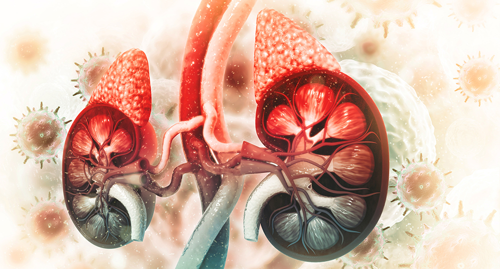
Two gene variants were studied in large-scale cohorts for their potential roles in bladder and kidney cancer among Polish patients.
Genitourinary cancers are a group of cancers that affect components of the urinary tract, including the bladder and kidneys. Worldwide, bladder and kidney cancer impact men at disproportionately higher rates than women. While incidence and mortality rates of bladder cancer in most western European countries have been consistently decreasing, some countries in the region, such as Poland, have seen an increase. Bladder cancer is the 4th most common malignancy in Polish men and the 14th most common malignancy in Polish women. There is currently a need to identify more effective bladder cancer biomarkers and therapeutic targets to develop new effective treatments that improve patient outcomes.
“The association between the NOD2 c.3020insC allele and CDKN2A missense variant c.442G>A (p.P.A148T) and survival of patients with bladder or kidney cancer remains controversial.”
In this study, the researchers investigated two gene variants—the NOD2 c.3020insC variant and the CDKN2A p.A148T polymorphism—and their role in bladder and kidney cancer in Polish cohorts. This NOD2 variant has been shown to occur in 7.3% of the Polish population. The CDKN2A polymorphism has been found in 3.5% of the Polish population. Therefore, these gene variants could be considered genetic risk factors for cancer. To test this hypothesis, the researchers assembled detailed participant data from a cohort of 706 bladder cancer patients and 410 kidney cancer patients. The team compiled control data from over 5,000 unselected, cancer-free individuals.
“To our knowledge, this is the first larger-scale study describing the clinical characteristics and survival of bladder and kidney cancer patients that is associated with the NOD2 c.3020insC allele and the CDKN2A p.A148T polymorphism in Poland.”
After performing the variant analysis in the cohort of Polish patients with bladder cancer, the team found that 8.9% of these patients carried the NOD2 variant and 5.2% carried the CDKN2A variant. However, their analysis revealed that neither the NOD2 nor the CDKN2A variant played a significant role in the survival of patients with bladder cancer. In performing the variant analysis in the cohort of Polish patients with kidney cancer, they found that 7.3% of these patients carried the NOD2 variant and 3.4% carried the CDKN2A variant. The researchers did not observe any statistically significant relationship between kidney cancer and either variant. However, they were not able to perform a survival analysis in the kidney cancer cohort.
Conclusion
The researchers found that the NOD2 c.3020insC variant and the CDKN2A p.A148T polymorphism were not significantly associated with the survival of bladder cancer patients, regardless of age, cancer family history, smoking status, and sex. To date, this is the first larger-scale study to examine these variants in association with clinical characteristics and survival of Polish patients with bladder cancer.
“In summary, the results of this study indicate that neither the NOD2 c.3020insC variant or the CDKN2A p.A148T polymorphism are associated with the survival of bladder cancer patients regardless of age, cancer family history, smoking status, and sex. Thus, the NOD2 c.3020insC or theCDKN2A p.A148T polymorphism cannot be added to the list of genes that are associated with an increased susceptibility to bladder or kidney cancer at this time.”
Click hereto read the full research paper published by Oncotarget.
Oncotarget is an open-access journal that publishes primarily oncology-focused research papers in a continuous publishing format. These papers are available at no cost to readers on Oncotarget.com. Open-access journals have the power to benefit humanity from the inside out by rapidly disseminating information that may be freely shared with researchers, colleagues, family, and friends around the world.
In a review paper published in Oncotarget in 2021, researchers discuss the impact of iron chelation on cancer cell survival and the underlying mechanisms of action.
Raw iron ore
Listen to an audio version of this article
Iron is essential for human life, however, this element can also become toxic in high doses. Contrary to iron anemia, iron overload occurs when the body accumulates more iron than it can use, and this excess iron is damaging to cells and tissues. Famous for their atypical growth patterns, cancer cells accumulate a surplus of iron to support their irregular growth and metabolism. Thus, the cancer-cell metabolism may be exploited by targeting their proclivity to require and retain iron.
“Iron chelators selectively deplete cancer cells of iron, exploiting cancer’s iron addiction – a trait displayed by a range of different cancers.”
Iron chelators are compounds that can bind to iron and facilitate iron wasting. Depriving cancer cells of iron using iron chelators has selectively cytotoxic effects in cancer cells. Some natural iron chelators include turmeric, quercetin, resveratrol, and green tea. Synthetic iron chelators include derivatives of 8-hydroxyquinoline, tachpyridine and deferoxamine. A considerable number of studies have shown that iron chelators can reverse some major catalysts and hallmarks of cancer—making iron chelators a promising treatment option for cancer patients.
Researchers Gina Abdelaal and Stephany Veuger from Northumbria University reviewed the available research literature about the impact of iron chelation on cancer cell survival and the underlying mechanisms of action. Their review paper was published by Oncotarget in 2021 and entitled, “Reversing oncogenic transformation with iron chelation.”
“This review aims to explore the underlying mechanisms of action behind iron chelator driven cytotoxicity in the context of the hallmarks of cancer established by Hanahan and Weinberg [47, 48] (see Figure 1, Supplementary Table 1). This will in turn support further research into iron chelators as a potential effective anti-cancer therapy.”
Iron Chelation Therapy
In the researchers’ review, they emphasize that iron chelation therapy has been shown to reverse multiple oncogenic hallmarks and is a promising treatment for many cancers. Studies have shown that iron chelation weakens cancer cell proliferation, induces cell cycle arrest, reactivates tumor suppressor genes, induces apoptotic signaling, inhibits stemness and Wnt/β-catenin signaling, prevents the initiation of metastasis through EMT and ROCK/MLC2 and NF-kB inhibition, and exploits and mimics genomic instability. While iron chelation has multiple targets within a cancer cell, the authors note that the NDRG1 gene has a critical role in inducing iron chelator-mediated cytotoxicity.
Figure 1: The impact of iron chelators on the hallmarks of cancer. Iron chelators have been shown to reverse many oncogenic signalling pathways associated with each hallmark of cancer with NDRG1 being a common thread. Generated through BioRender.com [47, 48].
One of the main mechanisms by which iron chelators exert their cytotoxic effects is through their ability to induce autophagy. However, this effect may both suppress and facilitate tumorigenesis. The researchers wrote that further in vivo studies must be conducted to reach a consensus about the impact of iron chelation on angiogenesis.
“In cancer cells, autophagy suppresses tumorigenesis by inhibiting cancer-cell survival and inducing cell death, but it also facilitates tumorigenesis by promoting cancer-cell proliferation and tumor growth [8,9].”
Many natural and synthetic iron chelators are currently being researched and developed. However, some early-developed iron chelators, such as deferoxamine, are effective in only some cancer patients. This is due to deferoxamine having poor lipophilicity, rapid clearance by the kidneys and poor absorption in the small intestine. Other iron chelators, such as those in the thiosemicarbazone class, are capable of inducing reactive oxygen species, causing oxidative stress. However, these chelators have only been successful in blood cancers, not in solid tumors. The researchers also spotlighted a novel iron chelator—VLX600—for its ability to target oxidative phosphorylation and initiate metabolic reprogramming.
“Cancer cells undergo a metabolic transformation known as the Warburg effect, which shifts their source of energy from oxidative phosphorylation to glycolysis. This is another trait which is exploited by iron chelators. VLX600 diminishes the ability of MCF7 and HCT116 cells to undergo oxidative phosphorylation [38].”
Conclusion
“Based on the data presented in this review iron chelators could potentially reverse many of the key hallmarks of cancer. Stripping the cells of iron impacts many cellular targets with some targets still undiscovered.”
The authors point out that the full impact of iron chelators on two remaining hallmarks of cancer, inflammation and immune evasion, have yet to be established. Additionally, the ability of iron chelators to induce both a pro-survival and tumor suppressor response in cancer cells through autophagy must be addressed. The researchers suggest that combining iron chelators with other inhibitors may be worth examination.
“We propose a combinatorial study of iron chelators with immune checkpoint inhibitors as they have shown success in clinic and could uncover more mechanisms of action.”
Click here to read the full review paper published in Oncotarget.
Oncotarget is a unique platform designed to house scientific studies in a journal format that is available for anyone to read without a paywall making access more difficult. This means information that has the potential to benefit our societies from the inside out can be shared with friends, neighbors, colleagues, and other researchers, far and wide.
Researchers demonstrated the overexpression of the protein APOBEC3B in adrenocortical carcinoma. They also identified the transcription factor, known as GATA3, that directly regulates APOBEC3B.
Listen to an audio version of this article
Adrenocortical carcinoma (ACC) is a rare and aggressive cancer that forms in the outer layer of the adrenal gland tissue above the kidneys. According to the National Institutes of Health, the occurrence of ACC in the United States is believed to only affect one to two people per million, per year. This highly-rare disease also challenges patients and researchers due to its post-diagnosis five-year survival rate of a mere 51%.
At this time, there are no known external factors that cause this disease. Most adrenocortical tumors that have been found produce symptoms including abdominal pain and higher levels of certain hormones, inclusive of cortisol, aldosterone, testosterone, and estrogen. Any of these hormones produced in excess can have numerous troubling effects on the body and, most alarmingly, the cancer cells in the adrenal glands have the potential to travel to other organs.
Previously, recent evidence confirmed the overexpression of a protein that is rightfully abbreviated as APOBEC3B (fully known as Apolipoprotein B mRNA editing enzyme catalytic subunit 3B) as a source of mutations occurring in breast, bladder, cervical, lung, head, and neck cancers. In this study the researchers used two publicly available datasets to analyze APOBEC3B gene expression in 21 normal adrenal cortices, 69 benign adrenocortical tumors and 38 ACC samples. They found that APOBEC3B is significantly overexpressed in ACC. The effects of this overexpression, in addition to a tumor mass, were consequently associated with DNA damage, reduced number of cells in S-phase arrest and increased alterations and gene mutations (particularly in the TP53 gene).
To assess the association between APOBEC3B and adrenocortical tumor growth, the team used mouse models to perform a “knockdown” or reduction, in APOBEC3B and measured the effects this had on the tumor tissue. The mice were divided into three groups of eight, and at weeks six and eight of the APOBEC3B knockdown, the researchers found significantly reduced cell proliferation and more cells in S-phase arrest.
GATA3 and APOBEC3B In ACC
The team was able to successfully knockdown APOBEC3B in mice and demonstrated that this caused a significant reduction in tumor volume. They also found in their analysis that tumors with higher expressions of APOBEC3B presented with a higher number of TP53 gene mutations. Given that the researchers were now confident that APOBEC3B is the protein that coincides with the growth of tumors in ACC, they sought to identify the mechanism responsible for regulating this protein.
After a thorough process of tests distinguishing between 90 different cancer-associated transcription factors, the team observed that the transcription factor GATA3 directly binds to the promoter region of APOBEC3B and transcriptionally regulates its gene expression in ACC.
Conclusion
In this study, the team successfully demonstrates that the protein APOBEC3B is overexpressed in ACC and causes DNA damage, alterations and mutations, and also, for the first time, that GATA3 directly regulates the expression of APOBEC3B. This confirms that the higher expression levels of both APOBEC3B and GATA3 are prognostic markers for patients with ACC.
This new information may be used in further research to develop treatments and interventions to improve the prognosis for those affected by adrenocortical carcinoma and other related disorders.
Click here to read the full research paper published by Oncotarget.
Oncotarget is a unique platform designed to house scientific studies in a journal format that is available for anyone to read without a paywall making access more difficult. This means information that has the potential to benefit our societies from the inside out can be shared with friends, neighbors, colleagues, and other researchers, far and wide.
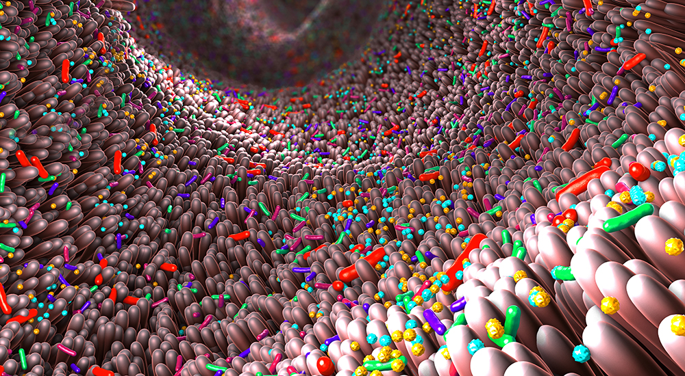
In this 2019 study, researchers investigated the effects of purified elements of cholera toxin in age-associated weight gain.
3D illustration of the gut microbiome
In recent years, scientists have made significant advancements to improve our understanding of the gut microbiome. This diverse environment—of somewhere around 39 trillion microorganisms living within the digestive tracts of vertebrates (including humans, and even insects)—includes bacteria, archaea, viruses, and fungi. However, a “healthy” gut microbiota remains difficult to define in humans. The contents of the gut microbiome are not only different between women and men, microbiomes differ between… everyone. Among unrelated humans, no more than 30% of the same bacterial strains are shared in the gut microbiome.
Different microbiomes can present with different biological reactions to outside factors, including infections and medications, and can even display different symptoms reacting to cancer and other diseases. Studies have repeatedly found that the gut microbiome plays important roles in human mood, sleep, metabolism, digestion, the immune and nervous systems, and in chronic inflammatory disorders, such as obesity.
“Indeed, earlier studies have shown that gut microbe-immune interactions contribute to smoldering inflammation, adiposity, and weight gain.”
The Hygiene Hypothesis
Researchers continue to find evidence to support the “hygiene hypothesis.” The hygiene hypothesis postulates that a lack of beneficial early-life microbe exposures can result in a dysregulated immune system later in life. This lack of early-life microbe exposures followed by immune imbalances may be responsible for the increase in obesity and other chronic inflammatory disorders over the past forty years.
“Systemic immune imbalances arising from the gut have been proposed as a probable cause of obesity [8].”
First, the researchers used both inbred and outbred mouse models to test the effects of the cholera-toxin subunit B (ctB)—a component of the Dukoral® vaccine used in humans for cholera diarrhea prevention. For each mouse model tested in the study, four different groups of eight mice each were examined: a female control group, a vaccinated female group, a male control group, and a vaccinated male group. At four weeks of age, the study mice were given three doses every-other-week of ctB at 10 micrograms. The control mice were given sham doses. The researchers found that in ctB vaccinated mice, the oral vaccination prevented age-associated weight gain compared to the control mice in both models.
Next, the researchers used an obese mouse model to test the effects of ctB dosing in early-life and to test the effects of transfering their gut flora into another mouse. The researchers found that the obese-mouse microbiome was sufficient to trigger obesity and inflammation in other mice when compared to sham-dosed control mice. In the obese mouse model, ctB dosing in early life also inhibited age-associated weight gain. This probiotic inhibited weight gain in mice dosed in early-life, and also in mice dosed in adulthood.
“Although we discovered dramatic benefit after early-life exposures to ctB, mice were also significantly slimmer when dosed with ctB for the first time during adulthood at 12-wks-of-age or 24-wks-of-age.”
Conclusion
The researchers found that purified elements of the cholera toxin stabilized immunity, through the gut microbiome, and inhibited age-associated obesity in multiple mouse models. Further studies are necessary to determine the degree to which an early-life microbe exposure such as this impacts immunity versus first-time adulthood exposures. Humans have been taking pre- and probiotics for quite some time without a strong grasp of exactly how these microbe infusions work. This research contributed to a better understanding of how humans can modulate our own gut microbiome to improve many aspects of our health and well-being.
“This type of microbe-immune re-programming may ultimately target other diseases linked with obesity and inflammation such as diabetes [19], multiple sclerosis [64], and cancer [25].”
Click here to read the full research paper, published by Oncotarget.
Oncotarget is a unique platform designed to house scientific studies in a journal format that is available for anyone to read without a paywall making access more difficult. This means information that has the potential to benefit our societies from the inside out can be shared with friends, neighbors, colleagues, and other researchers, far and wide.
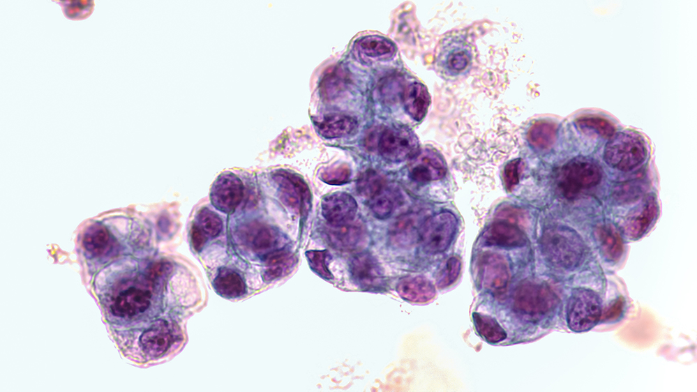
Researchers explored metformin with or without rapamycin as maintenance therapy in patients with metastatic pancreatic adenocarcinoma.
Malignant cells of adenocarcinoma
Maintenance chemotherapy has previously been recommended for patients with metastatic pancreatic ductal adenocarcinoma (mPDA)—as PDA is an aggressive cancer at all stages, and treatment options are limited for later-stage mPDA. However, maintenance chemotherapy regimens often lead to toxicity and are not viable long-term options. Therefore, researchers are exploring alternative maintenance therapies for mPDA patients. In preclinical studies, the therapeutic combination of metformin and rapamycin demonstrated a potential synergy of anti-tumor activity in PDA.
“A synergistic effect of the combination of metformin with rapamycin was suggested by preclinical studies demonstrating enhanced inhibition of mTOR in a pancreatic cancer cell line and better growth inhibition of pancreatic cancer cells in a xenograft tumor model with the combination than either agent alone [21].”
Metformin is an antihyperglycemic drug that is frequently prescribed for patients with diabetes to help control blood sugar levels. Rapamycin is an immunosuppressive drug that has historically been prescribed to prevent organ rejection in kidney transplant patients. (Today, rapamycin is also being considered for its potential use in anti-aging and longevity interventions.) In animals, metformin and rapamycin both inhibit the major biological regulator of growth, named the mammalian target of rapamycin (mTOR). mTOR is thought to be a main driver of many (if not all) aging-related diseases, including cancers such as PDA.
“Mechanistic/mammalian target of rapamycin (mTOR) is a serine/threonine protein kinase which acts as a signaling node downstream of several oncogenic pathways including KRAS/MEK/ERK and PI3K/Akt, both of which are thought to be relevant drivers in a majority of PDAs [6–9].”
A total of 22 unselected patients with mPDA were included in this randomized open-label phase 1b study between June 2014 and December 2017. Patients were at least 18 years of age and had previously been treated with chemotherapy for mPDA. At the beginning of the study, patients had either stable mPDA or responding mPDA for at least six months after induction chemotherapy. Half of the patients were randomly assigned to study Arm A, and the other 11 patients were assigned to study Arm B. Of note, the average age of the participants in Arm B was older (52–72; 66) than the participants in Arm A (34-73; 58). Otherwise, baseline characteristics between the study groups were relatively well-balanced.
Participants in study Arm A were assigned to take 850 milligrams of metformin orally, two times per day, for at least 12 months. Participants in study Arm B were assigned to take metformin and four milligrams of rapamycin once per day, for at least 12 months. The researchers conducted PET/CT scans, immunologic and metabolic analyses, statistical analysis, and continuously recorded and monitored for safety, patient tolerance, toxicity, and treatment-related adverse events.
“Treatment was continued until disease progression, intolerance of study treatments, or study closure, which occurred only after all remaining patients received a minimum of 12 months of treatment.”
Results and Conclusion
“In conclusion, the administration of metformin with or without rapamycin in patients with mPDA who achieve a response to chemotherapy is well-tolerated and was associated with better than expected overall survival in this study.”
The researchers observed “remarkably longer than expected” progression free survival and overall survival in this typically poor-prognosis population of patients. In this cohort, a low neutrophil-to-lymphocyte ratio and decreased fluorodeoxyglucose-avidity and/or decreased CA19-9 from baseline predicted improved outcomes among the long-term survivors. Overall, metformin and rapamycin were well-tolerated and their safety profiles were found to be comparable to previous reports. The researchers were forthcoming about limitations of their study—as their cohort was relatively small and the study was not powered to detect differences in clinical activity between the treatment arms.
“To this end, we identified several factors which may be used to select for patients with improved outcomes; however, whether good prognosis patients need any further treatment at all and whether poor prognosis patients will benefit from continued chemotherapy rather than a maintenance approach are not known and additional prospective studies are needed to answer these questions.”
Click here to read the full priority research paper, published by Oncotarget.
Oncotarget is a unique platform designed to house scientific studies in a journal format that is available for anyone to read without a paywall making access more difficult. This means information that has the potential to benefit our societies from the inside out can be shared with friends, neighbors, colleagues, and other researchers, far and wide.
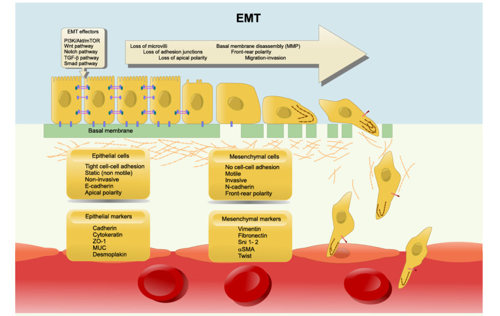
Researchers used mathematical modeling to investigate mechanisms that drive the elusive phenomenon of cancer cell resistance to epithelial-mesenchymal transition (EMT).
Epithelial–mesenchymal transition (EMT): losing cell polarity and cell adhesion to gain migratory and invasive properties.
Listen to an audio version of this article
Cancer cells have been known to use sagacious methods of evading apoptosis and mysteriously overcoming powerful anti-cancer therapies. One such method of evasion has recently been identified as the process of epithelial-mesenchymal transition (EMT) and its reverse process, mesenchymal-epithelial transition (MET). These transitions enable epithelial cells (structural/fixed) to gain mesenchymal cell (differentiating/mobile) functions, and viceversa. Researchers believe that epithelial-mesenchymal plasticity (EMP) allows cancers to become therapy resistant, determines cancer aggressiveness and allows metastatic cancer to mobilize and spread.
“Such dynamic and reversible switching can help tumor cells to overcome various challenges during disease progression such as anoikis [6], and assaults by the immune system [7].”
In the introduction of this paper, the authors discuss results from previous research about the reversibility and irreversibility of EMT/MET. EMT can be triggered by various EMT-inducing external signals, such as TGFβ or by adjusting the levels of EMT-specific transcription factors (EMT-TFs). They report that, in cells stimulated over shorter durations (between two and six days), cells may revert back to an epithelial state after withdrawal of the signal/stimulus. They also explain that cells that have been stimulated over longer durations (10+ days) may render EMT irreversible and to become “locked” in a mesenchymal state.
Researchers suspect the existence of a “tipping point” after continued signal/stimulus exposure is what results in irreversible EMT. Multiple mechanisms have been proposed as responsible for this tipping point, including epigenetic alterations and self-stabilizing feedback loops in regulatory circuits. However, there remains a need for studies to investigate the mechanistic basis that causes epithelial cells to be resistant to undergoing EMT, or the irreversibility of MET.
“Some sporadic observations about the resistance of epithelial cells to undergo EMT have been reported [14, 24], but a causative mechanistic understanding still remains elusive.”
The Study
To investigate the mechanisms that enable the irreversibility of MET, or lack of EMP, the researchers in this study used mechanism-based mathematical modeling. Their experimental observations indicated that a global epigenetic program limiting the action of ZEB1 was found to underlie epithelial trait retention in cells exposed to persistent Twist1 activation for 21 days. They demonstrated a possible underlying mechanism by which GRHL2 overexpression can resist EMT. Importantly, the researchers found that, from a single isogenic cell population, two subpopulations of cells emerged and responded differently to the EMT-signalling.
“Here, we propose two independent mechanism[s] that may explain the resistance of epithelial tumor cells to undergo EMT: 1) epigenetic feedback mediated via GRHL2—an MET-inducing transcription factor (MET-TF) [25–27]; and 2) stochastic partitioning of parent cell biomolecules among the daughter cells at the time of cell division [28–30].”
Aside from epigenetic mediation involving GRHL2, the researchers believe varying EMT-signal responses within isogenic cell populations are caused by stochastic partitioning of molecules during cell division. The researcher described this phenomenon as a type of incongruent “noise” that takes place when cells divide.
“Such noise in the distribution of molecules may affect cell-fate and drive non-genetic heterogeneity [28–30], leading to different phenotypic distributions in terms of EMT [3].”
Conclusion
The team concluded that MET should not only be considered the reverse process of EMT, as important and distinct processes may be involved in both EMT and MET transformations. The authors are forthcoming about limitations in their study—indicating that a more detailed molecular mechanism-based epigenetic model would provide better insights into EMT. They also note that they did not consider spatial effects in their model, where more dense or spread out cell populations and access to signal strength, nutrients and oxygen may change outcomes.
“Future efforts should decode the molecular mechanisms of any such epigenetic feedback of GRHL2 on ZEB1 expression as well as track the distribution of molecules during cell divisions happening while cells are being induced to undergo EMT/MET.”
Click here to read the full research paper published by Oncotarget.
Watch, read or listen to an Oncotarget Interview with Drs. Herbert Levine and Mohit Kumar Jolly as they discuss this paper.
Oncotarget is a unique platform designed to house scientific studies in a journal format that is available for anyone to read—without a paywall making access more difficult. This means information that has the potential to benefit our societies from the inside out can be shared with friends, neighbors, colleagues, and other researchers, far and wide.


![Figure 1: The impact of iron chelators on the hallmarks of cancer. Iron chelators have been shown to reverse many oncogenic signalling pathways associated with each hallmark of cancer with NDRG1 being a common thread. Generated through BioRender.com [47, 48].](https://www.impactjournals.com/wp-content/uploads/2022/02/Screen-Shot-2022-02-09-at-8.58.35-AM-999x1024.png)