In this new study, researchers present the first retrospective study evaluating differential gene expression of MIF, DDT, and relevant pathway markers in relation to clinical outcomes in melanoma patients.
—
Macrophage Migration Inhibitory Factor (MIF) and its homolog D-dopachrome Tautomerase (DDT) have been implicated as drivers of tumor progression in various cancers. Recent evidence suggests that MIF could be a therapeutic target in immune checkpoint inhibition (ICI) resistant melanomas; however, clinical evidence for MIF, and particularly for DDT, remains limited.
In their study, the researchers noted that melanoma is one of the most aggressive and lethal forms of cancer, with an estimated 99,700 new cases expected in 2024. The development of immune checkpoint inhibitors (ICIs) has significantly transformed cancer treatment and is now a cornerstone for managing several cancers, including advanced melanoma. Anti-CTLA-4 inhibitors, which target regulatory T cells, and anti-PD-1/L-1 inhibitors, which target activated T cells, dendritic cells, and tumor cells, have reshaped melanoma management, leading to improvements in progression-free and overall survival, with up to 22% of patients experiencing a complete response (CR). Data suggests that the ratio of CD74:MIF and CD74:DDT expression in melanoma may provide prognostic value and potentially serve as clinical biomarkers for patients with melanoma.
The study significantly expands on previous research by including a larger cohort of individuals and employing a comprehensive approach to defining high and low MIF and DDT expression. The survival analysis findings are consistent with existing literature, demonstrating that increased MIF levels are associated with worse prognosis in patients with melanoma, particularly in those with advanced disease or evidence of metastases.
The data presented in this research paper supports existing evidence on the intratumoral effects of MIF and DDT on tumor permissiveness, primarily through immune modulation, with implications for melanoma prognosis. The findings suggest that MIF and DDT may serve as therapeutic targets and biomarkers for predicting treatment response and survival, with CD74:MIF and CD74:DDT showing promise as markers of ICI response in patients undergoing treatment. Further investigation is needed to fully understand the role and functions of DDT in the melanoma microenvironment, as well as its distinct, non-overlapping functions in tumorigenesis.
“Our study is the first to report survival findings in association with intratumor DDT expression and CD74:DDT expression level ratio.” Click here to read the full research paper in Oncotarget.
—
Oncotarget is an open-access, peer-reviewed journal that has published primarily oncology-focused research papers since 2010. These papers are available to readers (at no cost and free of subscription barriers) in a continuous publishing format at Oncotarget.com.
Oncotarget is indexed and archived by PubMed/Medline, PubMed Central, Scopus, EMBASE, META (Chan Zuckerberg Initiative) (2018-2022), and Dimensions (Digital Science).
Click here to subscribe to Oncotarget publication updates.
This article dissects Dr. Mikhail V. Blagosklonny’s paradigm-shifting perspective on preemptive combinations for treating EGFR-mutant non-small cell lung cancer.
—
In the relentless battle against non-small cell lung cancer (NSCLC) driven by epidermal growth factor receptor (EGFR) mutations, the development of resistance has long been a formidable obstacle. Historically, first- and second-generation EGFR tyrosine kinase inhibitors (TKIs) like gefitinib, erlotinib, afatinib, and dacomitinib have faced a significant hurdle: the emergence of the T790M point mutation in approximately 50% of patients, rendering the tumor resistant to these therapies.
This resistance stems from a sobering reality – before treatment, a small subset of cancer cells already harbor the T790M mutation, conferring no selective advantage initially. However, once treatment commences, these rare mutated cells proliferate selectively, eventually dominating the tumor population and diminishing the effectiveness of first- and second-generation TKIs.
The Rise of Osimertinib: A Beacon of Hope
In 2015, the FDA approved osimertinib, a third-generation EGFR TKI, as a second-line therapy for NSCLC patients with the T790M mutation. This approval recognized that untreated tumors are typically T790M-negative, with the mutation potentially present in only a single cell initially. Moreover, approximately 50% of patients do not harbor this mutation at all, underscoring the rationale for administering osimertinib after resistance to first- or second-generation TKIs emerges.
However, a paradigm shift was on the horizon. Osimertinib’s ability to target the T790M mutation, coupled with its potential to eliminate the rare resistant cells before they proliferate, paved the way for a groundbreaking approach: administering osimertinib as a first-line treatment, without waiting for resistance to develop.
Preemptive Combinations: A Multifaceted Strategy
In a seminal 2018 clinical trial, osimertinib demonstrated its prowess as a first-line treatment, significantly extending median progression-free survival (PFS) compared to first-generation EGFR inhibitors (18.9 months vs. 10.2 months). Remarkably, while the objective response rates were similar between the two groups, the duration of response was nearly doubled with osimertinib (17.2 months vs. 8.5 months).
Capitalizing on these findings, Dr. Mikhail V. Blagosklonny introduced the concept of “preemptive combinations” – a multi-pronged approach to not only induce a therapeutic response but also eliminate the few resistant cells harboring pre-existing mutations. By combining osimertinib with first- or second-generation EGFR inhibitors like gefitinib and afatinib, these preemptive combinations could potentially prevent on-target resistance mechanisms, thereby extending PFS and overall survival (OS) for a substantial proportion of patients. On March 15, 2024, Dr. Blagosklonny’s research perspective was published in Oncotarget’s Volume 15, entitled, “From osimertinib to preemptive combinations.”
Expanding the Armamentarium: Comprehensive Preemptive Combinations
While osimertinib addresses the T790M mutation, other resistance mechanisms remain a concern. Approximately 50% of resistance cases involve on-target secondary mutations within EGFR, such as L718, G724, L792, G796, and C797, which can be targeted by first- or second-generation EGFR inhibitors. Additionally, off-target mechanisms like MET and HER2 amplifications contribute to resistance.
To combat this multifaceted challenge, Dr. Blagosklonny proposed a comprehensive preemptive combination comprising osimertinib, afatinib (or a first-generation EGFR inhibitor), and capmatinib (a potent MET inhibitor). This triple-threat approach could potentially prevent up to 75% of resistance mechanisms, dramatically extending median PFS from 18 months with osimertinib alone to an estimated 40 months.
Addressing Heterogeneity: The Bittersweet Reality
While osimertinib undoubtedly improves median PFS and OS compared to first-generation TKIs, a sobering reality emerges: approximately 20% of patients may experience a shortened PFS due to pre-existing mutations like C797S, which render the tumor resistant to osimertinib but sensitive to first-generation TKIs. In these cases, osimertinib inadvertently selects for resistant clones, potentially harming a subset of patients.
The solution, as proposed by Dr. Blagosklonny, lies in the simplicity of preemptive combinations. By combining osimertinib with gefitinib, or even a triple combination with afatinib, the risk of selecting for resistant clones is mitigated, ensuring that no patient is inadvertently harmed by the superior TKI.
Transient Combinations: A Flexible Approach
For clinicians who may be hesitant about administering three- or four-drug combinations, Dr. Blagosklonny suggests a flexible approach: a sequence of transient two-drug combinations. This strategy involves alternating between different combinations, such as osimertinib and gefitinib, followed by osimertinib and afatinib, and so on, effectively covering all potential resistance mechanisms while maintaining a manageable treatment regimen.
Extending the Paradigm: MET-Driven NSCLC
The insights gleaned from EGFR-driven NSCLC can be applied to other molecular subtypes, such as MET exon 14 skipping mutation (METex14)-driven NSCLC. Dr. Blagosklonny’s personal experience with this condition underscores the importance of preemptive combinations in this setting as well.
While the selective MET inhibitor capmatinib demonstrated remarkable efficacy in treating Dr. Blagosklonny’s brain metastases, resistance tends to emerge within a year, often driven by secondary mutations like D1228 and Y1230. To combat this, a preemptive combination of capmatinib, afatinib (to target EGFR and HER2 alterations), and cabozantinib (a type II MET inhibitor effective against resistance mutations) could potentially prevent up to 50% of resistance mechanisms, prolonging progression-free survival for a significant proportion of METex14-driven NSCLC patients.
Overcoming Hurdles: The Path Forward
Despite the promise of preemptive combinations, challenges remain. The development of novel targeted therapies and the exploration of immune checkpoint inhibitors in combination with these regimens offer exciting avenues for future research. Additionally, refining clinical trial designs to incorporate precision medicine approaches and tailored combination strategies will be crucial in translating these concepts into tangible benefits for patients.
Conclusion: A Paradigm Shift in Cancer Care
Dr. Blagosklonny’s perspective on preemptive combinations represents a paradigm shift in the treatment of lung cancer and potentially other malignancies. By proactively targeting resistance mechanisms before they can take hold, this approach offers a glimmer of hope for prolonging progression-free survival and improving outcomes for patients grappling with this disease. As research continues to unravel the complexities of cancer resistance, preemptive combinations may pave the way for a future where we can stay one step ahead of cancer.
Click here to read the full research perspective in Oncotarget.
—
Oncotarget is an open-access, peer-reviewed journal that publishes primarily oncology-focused research papers. These papers are available to readers (at no cost and free of subscription barriers) in an open-access and continuous publishing format at Oncotarget.com.
Oncotarget is indexed and archived by PubMed/Medline, PubMed Central, Scopus, EMBASE, META (Chan Zuckerberg Initiative) (2018-2022), and Dimensions (Digital Science).
Click here to subscribe to Oncotarget publication updates.
In a new study, researchers investigated the activity of gartisertib, a potent ATR inhibitor, alone and in combination with standard therapy in multiple glioblastoma cell lines.
—
Glioblastoma is a type of brain cancer that is very aggressive and difficult to treat. The current standard treatment involves surgery, radiation therapy, and chemotherapy with a drug called temozolomide (TMZ). However, many glioblastoma cells can resist the DNA-damaging effects of TMZ and radiation by activating a mechanism called the DNA damage response (DDR). This mechanism, while beneficial in normal cells, is detrimental to cancer therapy because it allows cancer cells to repair damage and continue to grow and divide. There is a need to counteract this mechanism in glioblastoma cancer cells.
“Here, we investigated the activity of gartisertib, a potent ATR inhibitor, alone and in combination with TMZ and/or RT in 12 patient-derived glioblastoma cell lines.”
The Study
In this study, the team tested the effects of gartisertib alone and in combination with TMZ and radiation in 12 patient-derived glioblastoma cell lines. They found that gartisertib alone reduced the viability of glioblastoma cells, and that the sensitivity was associated with the frequency of DDR mutations and the expression of genes involved in the G2 phase of the cell cycle (the phase where cells prepare for division and check for DNA damage). The researchers also found that gartisertib enhanced the cell death induced by TMZ and radiation, and that the combination was more synergistic than TMZ and radiation alone.
Interestingly, gartisertib was more effective in glioblastoma cells that had unmethylated MGMT promoters and were resistant to TMZ and radiation. (MGMT is a gene that encodes an enzyme that can reverse the damage caused by TMZ, and its promoter is a region that controls its expression. Methylation is a chemical modification that can silence genes, so unmethylated MGMT promoters mean higher MGMT expression and more resistance to TMZ.) The researchers also analyzed the gene expression changes in glioblastoma cells treated with gartisertib, and found that the drug upregulated pathways related to the innate immune system. The researchers speculated that gartisertib may trigger an immune response against glioblastoma cells, which could enhance the anti-tumor effects of the drug.
“We showed that gartisertib alone potently reduced the cell viability of glioblastoma cell lines, where sensitivity was associated with the frequency of DDR mutations and higher expression of the G2 cell cycle pathway. ATR inhibition significantly enhanced cell death in combination with TMZ and RT and was shown to have higher synergy than TMZ+RT treatment. MGMT promoter unmethylated and TMZ+RT resistant glioblastoma cells were also more sensitive to gartisertib. Analysis of gene expression from gartisertib treated glioblastoma cells identified the upregulation of innate immune-related pathways.”
Conclusion
The study is the first to demonstrate the activity of gartisertib in patient-derived glioblastoma cell lines, and it provides evidence that ATR inhibition may be a promising strategy to improve the outcomes of glioblastoma patients. Gartisertib is a potent and selective inhibitor of ATR that has been tested in a phase 1 clinical trial for patients with advanced solid tumors. The researchers suggest that further studies are needed to evaluate the safety and efficacy of gartisertib in combination with TMZ and radiation in glioblastoma patients, and to explore the potential role of the immune system in mediating the anti-tumor effects of the drug.
“In conclusion, this study identifies gartisertib as a potent ATRi within patient-derived glioblastoma cell lines. […] Further investigation of the concept of ATR inhibition for treatment of brain tumours, especially in vivo with brain penetrant compounds, is needed to validate these findings. Lastly, ATR inhibition alters the gene expression of innate immune and inflammatory signalling pathways within glioblastoma cells, which requires additional validation and investigation as a strategy to provoke an immunomodulatory response.”
Click here to read the full research paper in Oncotarget.
—
Oncotarget is an open-access, peer-reviewed journal that has published primarily oncology-focused research papers since 2010. These papers are available to readers (at no cost and free of subscription barriers) in a continuous publishing format at Oncotarget.com. Oncotarget is indexed/archived on MEDLINE / PMC / PubMed.
Click here to subscribe to Oncotarget publication updates.
In this new study, researchers investigated the role of Osteopontin splice variants in cancer metastasis.
—
Mitochondrial biogenesis, the process of increasing the size and number of mitochondria within cells, plays a crucial role in cancer metastasis. Metastasizing cells exhibit a unique metabolism that differs from the well-known Warburg effect observed in primary tumors. While primary tumors primarily rely on glycolysis for energy production, metastatic cells rely on oxidative phosphorylation and ATP generation for short-term energy needs. However, over longer time frames, mitochondrial biogenesis becomes a prominent feature in the success of metastasis.
In a new study, researchers Gulimirerouzi Fnu and Georg F. Weber from the University of Cincinnati’sJames L. Winkle College of Pharmacy investigate the connection between short-term oxidative metabolism and long-term mitochondrial biogenesis in cancer metastasis. They hypothesized that Osteopontin splice variants, specifically Osteopontin-c, stimulate an increase in mitochondrial size through the activation of specific signaling mechanisms. On December 1, 2023, their new research paper was published in Oncotarget, entitled, “Osteopontin induces mitochondrial biogenesis in deadherent cancer cells.”
“Over longer time frames, mitochondrial biogenesis becomes a pronounced feature and aids metastatic success. It has not been known whether or how these two phenomena are connected. We hypothesized that Osteopontin splice variants, which synergize to increase ATP levels in deadherent cells, also increase the mitochondrial mass via the same signaling mechanisms.”
The Role of Osteopontin Variants in Mitochondrial Biogenesis
Deadhesion, the process of detaching cancer cells from the extracellular matrix, is known to induce metabolic reprogramming and promote cancer cell survival in circulation. Osteopontin (OPN), a cytokine produced by cancer cells, has been implicated in tumor progression and the development of metastases. It mediates tumor cell survival and expansion under deadherent conditions, making it an ideal candidate for studying the mechanisms behind mitochondrial biogenesis. The authors of the research paper focused on two Osteopontin splice variants, Osteopontin-a and Osteopontin-c, and their effects on mitochondrial biogenesis.
Through their experiments with breast tumor cells, the authors found that both Osteopontin-a and Osteopontin-c contribute to mitochondrial biogenesis in deadherent cells. However, Osteopontin-c was more effective in stimulating an increase in mitochondrial size compared to Osteopontin-a. The authors also observed that the autocrine effects of Osteopontin variants are critical for the survival and anchorage-independence of disseminating malignant cells.
The Role of CD44v and SLC7A11 in Osteopontin Signaling
To further elucidate the mechanism behind Osteopontin-induced mitochondrial biogenesis, the authors investigated the receptors involved in Osteopontin signaling. They focused on CD44, a cell surface receptor known to interact with Osteopontin, and its variant CD44v. The authors found that Osteopontin-induced mitochondrial biogenesis is mediated via the binding of Osteopontin to CD44v.
Additionally, the authors discovered that the chloride-dependent cystine-glutamate transporter SLC7A11 plays a crucial role in Osteopontin signaling. The upregulation and co-ligation of SLC7A11, along with CD44v, leads to the activation of PGC-1, a known inducer of mitochondrial biogenesis. Surprisingly, the authors found that peroxide, an important intermediate in this signaling cascade, acts upstream of PGC-1 and is likely produced as a consequence of SLC7A11 recruitment and activation.
In Vivo Implications and Therapeutic Targets
To validate the relevance of their findings in clinical settings, the authors analyzed gene expression profiles in breast cancer metastases and metastases from other types of cancers. They identified the master regulator of mitochondrial biogenesis, PPARG, as well as its downstream effectors NRF1 and BACH1, to be upregulated in various metastases. These findings suggest that the Osteopontin-induced activation of PGC-1 and subsequent mitochondrial biogenesis may play a crucial role in cancer metastasis.
The authors also conducted in vivo experiments using mouse models. They observed that suppression of the biogenesis-inducing mechanisms led to a reduction in disseminated tumor mass. These findings not only confirm the functional connection between short-term oxidative metabolism and long-term mitochondrial biogenesis in cancer metastasis but also provide potential mechanisms and targets for treating cancer metastasis.
Conclusion
This study provides valuable insights into the role of Osteopontin splice variants in regulating mitochondrial biogenesis in metastatic cancer cells. The researchers demonstrated that Osteopontin-c stimulates an increase in mitochondrial size through the activation of specific signaling mechanisms involving CD44v and SLC7A11. These findings have significant implications for understanding the metabolic adaptations of metastatic cancer cells and suggest potential targets for therapeutic interventions. Further research is needed to fully elucidate the intricate signaling pathways involved in Osteopontin-induced mitochondrial biogenesis and to explore the clinical applications of these findings in cancer treatment.
“This study confirms a functional connection between the short-term oxidative metabolism and the longer-term mitochondrial biogenesis in cancer metastasis – both are induced by Osteopontin-c. The results imply possible mechanisms and targets for treating cancer metastasis.”
Click here to read the full research paper in Oncotarget.
—
Oncotarget is an open-access, peer-reviewed journal that has published primarily oncology-focused research papers since 2010. These papers are available to readers (at no cost and free of subscription barriers) in a continuous publishing format at Oncotarget.com. Oncotarget is indexed/archived on MEDLINE / PMC / PubMed.
Click here to subscribe to Oncotarget publication updates.
In this editorial, researchers delve into the immunotherapeutic challenges posed by the tumor microenvironment and liver metastasis in pancreatic cancer.
—
Pancreatic ductal adenocarcinoma (PDA), a common type of pancreatic cancer, has proven to be largely resistant to immunotherapy, a treatment that uses the body’s immune system to fight cancer. Despite numerous successful pre-clinical trials using sophisticated PDA mouse models, clinical trials have failed to show a significant improvement in survival.
Tumor Microenvironment and Liver Metastasis: Challenges in Pancreatic Cancer
The authors attribute PDA immunotherapy resistance to the unique characteristics of the tumor microenvironment (TME). The TME is often hypoxic and fibrotic, making it inaccessible to immune cells. Furthermore, the immune cells that do infiltrate the TME often have tolerogenic features, meaning they are more likely to tolerate the presence of cancer cells rather than attack them.
PDA most commonly metastasizes to the liver, an organ known for its immune tolerance. The liver is home to a diverse array of innate immune populations, including NK cells, Kupfer cells, NKT cells, and double negative T cells. Despite this, the liver is the most common location for metastasis from gastrointestinal cancers.
“It is an unfortunate fact that all failed clinical trials assessing immunotherapeutic efficacy were conducted in metastatic PDA, whereas basic preclinical investigations are usually performed in primary PDA using genetically engineered mouse models. We postulated that this dichotomy may explain the gap between preclinical promise and ultimate clinical failure.”
Divergent Responses to Immunotherapy: Primary vs. Metastatic
“The potentially divergent responses to immunotherapy in the respective environments of primary versus metastatic PDA within the same host has not been well-studied.”
The authors highlight the lack of research into the potentially divergent responses to immunotherapy in primary versus metastatic PDA. They argue that this gap in knowledge may explain the discrepancy between the promising results of pre-clinical trials and the disappointing outcomes of clinical trials.
In their research, they discovered that the TMEs of primary PDA and liver metastases differ significantly, and this difference plays a critical role in the site-specific response to immunotherapy. They found that liver metastases are uniquely resistant to immunotherapies, in stark contrast to the immunotherapeutic responsiveness of primary PDA.
“We discovered that the respective TMEs of primary PDA and liver metastases differ markedly and this fact plays a critical role in dictating site-specific PDA response to immunotherapy [6].”
The Role of B Cells
The researchers identified B cells as a key player in this differential response. They found that B cells constituted approximately 25% of the tumor-infiltrating lymphocytes in metastatic PDA liver deposits, compared to approximately 10% in primary PDA. They also discovered a novel population of CD24+CD44–CD40– B cells in the metastatic liver, which is recruited to the metastatic milieu by Muc1hiIL18hi tumor cells.
“[…] by targeting B cells or blocking CD200/BTLA, we demonstrated enhanced macrophage and T-cell immunogenicity, which enabled immunotherapeutic efficacy of liver metastases.”
However, the authors note that primary PDA sites lack this b-cell population. Instead, they are characterized by macrophages and effector T cells that have a higher ability to provoke an immune response. This makes their immunotherapeutic responsiveness far more robust than metastatic liver PDA.
Conclusion
This research underscores the importance of understanding the immune basis of differential responses to immunotherapy in primary versus metastatic pancreatic cancer. It highlights the need for further research into the role of the TME and immune cells like B cells in the response to immunotherapy. Such insights could pave the way for more effective treatments for this challenging disease.
“[…] our data suggest that models of primary PDA are poor surrogates for evaluating immunity or treatment response in advanced disease.”
Click here to read the full editorial paper in Oncotarget.
—
Oncotarget is an open-access, peer-reviewed journal that has published primarily oncology-focused research papers since 2010. These papers are available to readers (at no cost and free of subscription barriers) in a continuous publishing format at Oncotarget.com. Oncotarget is indexed/archived on MEDLINE / PMC / PubMed.
Click here to subscribe to Oncotarget publication updates.
In a new editorial paper, researchers from Tel Aviv University discuss a recent study exploring how whole-genome doubling shapes the aneuploidy landscape of human cancers.
—
Whole-genome doubling (WGD) and aneuploidy are two common genomic alterations that occur in human cancers. WGD is a macro-evolutionary event that results in the duplication of the entire genome, while aneuploidy is a micro-evolutionary event that results in the gain or loss of individual chromosomes or chromosome arms. Both WGD and aneuploidy can have profound effects on cellular physiology, gene expression and genome stability, and are associated with tumor initiation, progression and drug resistance.
“It is known that tumors that have undergone WGD are more permissive to aneuploidy, but whether WGD also affects aneuploidy patterns has remained an open question.”
The Study
The researchers analyzed 5,586 clinical tumor samples that had not undergone WGD (WGD-) and 3,435 tumors that had (WGD+) from The Cancer Genome Atlas (TCGA), across 22 tumor types. They found that WGD+ tumors were characterized by more promiscuous aneuploidy patterns, in line with increased aneuploidy tolerance. The relative prevalence of recurrent aneuploidies decreased in WGD+ tumors, suggesting that WGD+ tumors are more tolerant to aneuploidy than WGD- tumors.
The genetic interactions between chromosome arms differed between WGD- and WGD+ tumors, resulting in different co-occurrence and mutual exclusivity patterns. The proportion of whole-chromosome aneuploidy was significantly higher in WGD+ tumors than in WGD- tumors, indicating that different mechanisms of aneuploidy formation are dominant in WGD- and WGD+ tumors. The authors proposed that whole-chromosome missegregation is more prevalent in WGD+ tumors due to increased centrosome amplification and multipolar mitoses.
To validate their findings from the clinical tumor analysis, the authors used human cancer cell lines that reproduced the WGD/aneuploidy interactions observed in vivo. They also induced WGD in human colon cancer cell lines by treating them with a microtubule-stabilizing drug, and followed the evolution of aneuploidy in the isogenic WGD+/WGD- cells under standard or selective conditions. These experiments confirmed that WGD alters the aneuploidy landscape of human cancer cells, and revealed a causal link between WGD and altered aneuploidy patterns.
“We note that these experiments were not powered to assess the associations between specific aneuploidies, which remain to be experimentally validated in future studies.”
Conclusions & Future Studies
In their editorial, the researchers note that their study prompts questions about how different tetraploidization methods affect aneuploidy landscapes. They used cytokinesis failure for cell lines, but processes like cell fusion could impact aneuploidy differently. Further research should explore how selection pressures shape karyotype evolution, considering factors beyond tissue type. Analyzing intra-chromosomal arm-level vs. whole-chromosomal aneuploidies may identify cancer-driving chromosome arms. Overall, this study provides novel insights into how WGD and aneuploidy interact in human cancer, and how this interaction affects tumor evolution. The authors suggest that the interaction between WGD and aneuploidy is a major contributor to tumor heterogeneity, adaptation, and drug resistance, and that targeting this interaction could be a promising therapeutic strategy.
“In summary, our recent study shows that WGD contributes to aneuploidy formation in human tumors in both qualitative and quantitative ways. Hence, we propose that the WGD status of the tumor should be taken into account when examining the tumorigenic role of individual aneuploidies or aneuploidy patterns. In general, WGD should be considered in the study of aneuploidy landscapes in human cancers.”
Click here to read the full editorial in Oncotarget.
—
Oncotarget is an open-access, peer-reviewed journal that has published primarily oncology-focused research papers since 2010. These papers are available to readers (at no cost and free of subscription barriers) in a continuous publishing format at Oncotarget.com. Oncotarget is indexed/archived on MEDLINE / PMC / PubMed.
Click here to subscribe to Oncotarget publication updates.
In a new review, researchers from The Hebrew University of Jerusalem discuss the challenges associated with targeting Ras proteins and how protein engineering has emerged as a promising method to overcome these challenges.
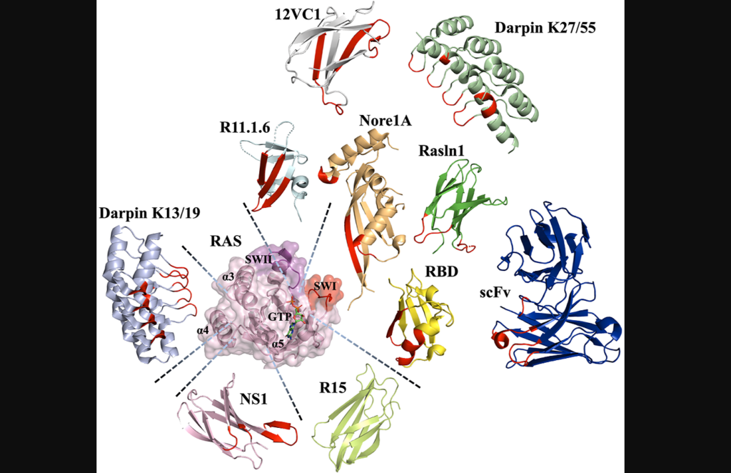
Figure 3: Various scaffolds utilized to engineer binders to Ras and their binding epitopes.
Ras plays a crucial role in controlling various cellular processes by switching between active (Ras-GTP) and inactive (Ras-GDP) states with the help of specific molecules. In its active form, Ras interacts with multiple effector proteins, initiating downstream events. Humans have three Ras genes, resulting in four isoforms that have distinct expression patterns and unique functions in different tissues. Posttranslational modifications target Ras to the cell membrane, where it can form dimers and interact with effectors through common domains. Ras mutations, commonly found in pancreatic, colorectal and lung cancers, lock Ras in an active state, promoting continuous cell division and proliferation. Ras signaling disruption occurs through reduced catalytic activity, altered effector binding and decreased affinity for other regulatory proteins.
Although Ras has been considered difficult to target, recent advancements have identified potential binding pockets that can be addressed by small molecules, peptidomimetics and proteins. Inhibitors designed to covalently bind to the Ras G12C mutant have shown promise, leading to FDA-approved drugs for specific lung cancers. Additionally, protein-based inhibitors that target Ras and its interactions with effectors, regulatory proteins and guanine nucleotide exchange factors offer alternative strategies for therapeutic intervention. These developments have challenged the notion that Ras is “undruggable” and highlight the potential for effective treatments against various cancer types.
On July 1, 2023, researchers Atilio Tomazini and Julia M. Shifman from The Hebrew University of Jerusalem published a new review paper in Oncotarget, entitled, “Targeting Ras with protein engineering.” The authors provide an overview of the challenges associated with targeting Ras proteins with small molecules and discuss how protein engineering has emerged as a promising method to overcome these challenges.
“While the development of small-molecule Ras inhibitors has been reviewed elsewhere [40], we focus our review on protein-based Ras inhibitors, describing the methods for their engineering, various scaffolds used for inhibitor design, and prospects for delivery of the designed Ras inhibitors into the cellular cytoplasm, where Ras is located.”
Protein Engineering
Protein scaffolds offer alternative approaches to small molecule drugs for engineering protein-based inhibitors. Unlike small molecules, protein domains can bind to targets through large surface areas, providing high affinity and specificity. Antibodies, natural protein effectors and novel binding domains are commonly used as protein scaffolds. Antibodies can be engineered into smaller versions to overcome limitations, while natural effectors can be modified to enhance binding affinity. Novel binding domains, unrelated to the target protein, possess structural robustness and can be evolved to exhibit strong binding. All three classes of protein scaffolds have been utilized to engineer Ras binders and explore strategies to inhibit Ras oncogenesis.
“Interestingly, all classes of protein scaffolds, including antibodies, natural effectors, and novel binding domains, have been utilized for engineering of Ras binders, allowing scientists to target various sites on the Ras surface and to explore different strategies for inhibiting Ras oncogenesis […].”
Methods for engineering protein inhibitors can be categorized into experimental directed evolution and computational design, or a combination of both. Experimental techniques involve display technologies such as phage display, yeast surface display, ribosome display, and mRNA display. These methods allow for the construction of combinatorial libraries of protein mutants, which are then screened using the target protein as a selection “bait.” The selected binders are sequenced to identify high-affinity mutants. Negative selection steps can be incorporated to enhance specificity by eliminating binders to unwanted targets. The number of mutants that can be assayed depends on the display technology used, with each approach having its limitations.
In addition to experimental approaches, computational methods have been proposed for protein binder design. Computational design enables rational targeting of specific binding epitopes on the target protein. However, computationally designed binders often have weak initial binding affinities and require affinity maturation through experimental techniques. Computational methods have been successful in designing focused libraries for yeast surface display experiments, where small libraries of protein mutants are designed based on computational predictions. This approach narrows down the choices to the most promising mutants, facilitating directed evolution experiments. By combining computational and experimental approaches, protein inhibitors with superior affinity and specificity have been developed.
“We have summarized all the described engineered Ras protein-based binders and their properties in Table 1.”
The Future of Intracellular Transport for Ras Inhibitors
Efficient delivery of molecules that bind to intracellular Ras proteins is essential for suppressing pro-cancer pathways and promoting anti-cancer activities. To overcome the challenge of crossing the cell membrane, different strategies have emerged. One approach involves utilizing short cell-penetrating peptides (CPPs) that can be fused to the desired protein, allowing entry into cells through direct translocation or endocytosis. However, improving the release of cargo proteins from endosomes remains a hurdle. Supercharging proteins with positively charged surfaces or leveraging bacterial toxins with intrinsic delivery mechanisms are alternative methods for intracellular protein delivery. Additionally, coupling cargo proteins to nanoparticles or employing mRNA delivery systems have shown promise, although they have their own limitations.
These protein delivery techniques have been explored for targeting Ras inhibitors. For instance, a human IgG1 antibody was engineered to selectively bind to Ras-GTP, inhibiting downstream signaling. Fusion of Ras binding domains to CPPs demonstrated competitive inhibition of Ras/effector interactions. Furthermore, optimized bacterial secretion systems and lipid nanoparticle-encapsulated mRNA platforms have been employed for efficient intracellular delivery of Ras-binding molecules. These advancements open up possibilities for targeted cancer therapies and disease treatments by enabling effective delivery of Ras binders to their intracellular target, thus influencing cancer-related signaling pathways.
Conclusions
In summary, targeting Ras proteins, despite their historically challenging nature, has seen significant progress in recent years. Small molecules, peptidomimetics and protein-based inhibitors have emerged as potential strategies for inhibiting Ras oncogenesis. Protein engineering, utilizing various protein scaffolds such as antibodies, natural effectors and novel binding domains, offers alternative approaches to traditional small molecule drugs.
Experimental directed evolution and computational design, alone or in combination, have facilitated the development of high-affinity and specific protein inhibitors. Furthermore, the efficient intracellular delivery methods described above hold promise for targeted cancer therapies by effectively delivering Ras binders to their intracellular targets. These advancements challenge the perception of Ras as “undruggable” and provide hope for the development of effective treatments for various cancer types.
“These strategies should be utilized in future to examine the beneficial activity of Ras-binders and inhibitors and should further facilitate the development of protein-based Ras therapeutics.”
Oncotarget is an open-access, peer-reviewed journal that has published primarily oncology-focused research papers since 2010. These papers are available to readers (at no cost and free of subscription barriers) in a continuous publishing format at Oncotarget.com. Oncotarget is indexed/archived on MEDLINE / PMC / PubMed.
Click here to subscribe to Oncotarget publication updates.
In a new editorial, researchers discuss a study using their team’s new genetically engineered mouse (GEM) model to assess PLK1 as a driver of oncogenic transformation.
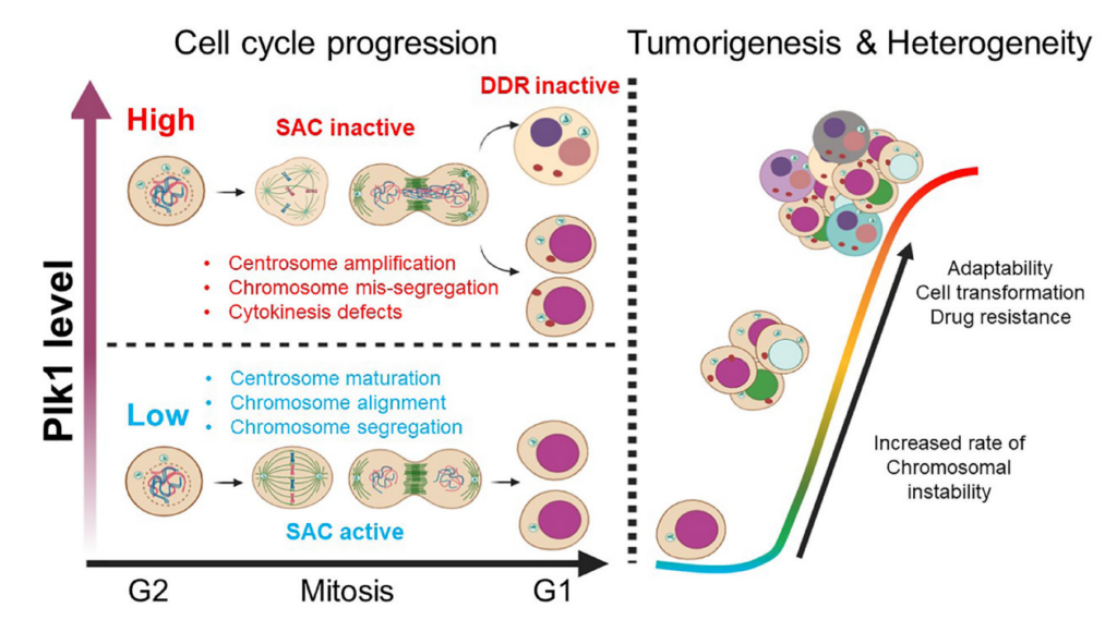
On the bright side, polo-like kinase 1 (PLK1) is considered a master regulator of the ever-important cell cycle. On the dark side, PLK1 expression (at both the mRNA and protein level) has shown to be upregulated in tumor cells, suggesting that PLK1 may also contribute to tumorigenesis. Despite this direct association, researchers studying the role of PLK1 in cancer have encountered a problem: a lack of appropriate animal models for experimentation.
“Even though studies have suggested that PLK1 contributes to tumorigenesis, the ability of PLK1 to drive oncogenic transformation on its own in vivo was still questionable due to a lack of sophisticated animal models for experimentation [18, 19].”
“To address this important scientific question, we generated a new genetically engineered mouse (GEM) model using the CAGGS (cytomegalovirus (CMV) early enhancer/chicken β-actin) promoter to drive exogenous PLK1 expression, allowing its ubiquitous and robust gene expression in transgenic mice [20].”
In an effort to determine if PLK1 overexpression causes tumors, the researchers created a new GEM mouse model that expresses high levels of PLK1. These high levels caused various types of spontaneous tumors. The increased PLK1 levels caused defects in cell division and resulted in abnormal numbers of centrosomes and compromised cell cycle checkpoints. This allowed for the accumulation of chromosomal instability, leading to abnormal numbers of chromosomes and tumor formation. In human cancers, higher PLK1 expression was associated with an increase in genome-wide copy number alterations. Their study provides evidence that abnormal PLK1 expression can trigger chromosomal instability and tumor formation, suggesting potential therapeutic opportunities for cancers with chromosomal instability.
“In summary, this study provides a novel GEM model that recapitulates the increased PLK1 expression observed in many human cancers and demonstrates that PLK1 overexpression drives spontaneous tumor formation in multiples organs in mouse, revealing the dark side of PLK1 as a potent proto-oncogene.”
Conclusions
In conclusion, the limitations of previous studies on PLK1 and its role in cancer have been partially addressed by the development of the new GEM model created by these researchers. This model allowed the team to examine PLK1’s ability to drive oncogenic transformation in vivo. Their study demonstrates that overexpression of PLK1 leads to the formation of spontaneous tumors in multiple organs, highlighting the dark side of PLK1 as a potent proto-oncogene. The findings of this study provide valuable insights into the role of PLK1 in tumorigenesis and suggest potential therapeutic opportunities for cancers associated with chromosomal instability. This breakthrough in animal models opens up new avenues for further research in understanding the mechanisms underlying PLK1-related tumorigenesis and developing targeted therapies to combat cancer.
“Alternative therapeutic strategies, such as co-delivery systems using nanoparticles or combination therapies, are under development in order to enhance the efficacy of PLK1 inhibition [25–28]. With expanding discoveries of PLK1 function and mechanisms of action, we hope that PLK1-targeted therapies will soon join the frontlines in the fight against cancer.”
Click here to read the full editorial paper in Oncotarget.
Oncotarget is an open-access, peer-reviewed journal that has published primarily oncology-focused research papers since 2010. These papers are available to readers (at no cost and free of subscription barriers) in a continuous publishing format at Oncotarget.com. Oncotarget is indexed/archived on MEDLINE / PMC / PubMed.
Click here to subscribe to Oncotarget publication updates.
In a new study, researchers found that 38.4% of a cohort in Saudi Arabia carried pathogenic variants linked to hereditary cancer risk.
Familial cancer is a fearsome reality for millions of people worldwide. While some cases of familial cancer syndrome (FCS) may be influenced by shared environmental or lifestyle factors within a family, others are solely due to genetic mutations passed down through generations. This problem is especially prevalent in Saudi Arabia—where rates of familial cancer are among the highest in the world.
“Cancer increased in the Kingdom of Saudi Arabia by 136% between 1999 and 2015 [4].”
Approximately 20% of all Saudi Arabian cancer patients have a family history of cancer. This population is likely to carry mutant alleles, presenting an opportunity for further exploration and research. By studying these individuals and their genetic profiles, scientists and healthcare professionals can gain valuable insights into the genetic factors contributing to familial cancer in the Saudi Arabian population. This knowledge can help improve risk assessment, develop targeted prevention strategies, and potentially lead to more effective treatments for familial cancer cases.
The researchers used a 30-gene, targeted NGS panel to screen 310 subjects, including 57 non-cancer patients, 110 index patients with cancer and 143 of their relatives, 16 of whom also had cancer. (“Index patients” refers to individuals who are the first in a family to be diagnosed with a particular disease or condition of interest.) The NGS panel covered genes related to breast, ovarian, colorectal, endometrial, gastric, pancreatic, prostate, thyroid, renal, and skin cancers, as well as familiar adenomatous polyposis (FAP) and Lynch syndrome.
“This kit has been previously trialed as a means of capturing potential PVs [pathogenic variants] at a population level in Nigeria and the Caribbean, and in identifying rare variants in cancer patients who have tested negative for common cancer variants [35–38].”
The results showed that 119 subjects (38.4% of the cohort) carried pathogenic or likely pathogenic variants (PVs) affecting genes associated with hereditary cancer risk. (TP53, ATM, CHEK2, CDH1, CDKN2A, BRCA1, BRCA2, PALB2, BRIP1, RAD51D, APC, MLH1, MSH2, MSH6, PMS2, PTEN, NBN/NBS1, and MUTYH were identified as genes with pathogenic or likely pathogenic variants.) Among 126 patients and relatives with a history of cancer, 49 subjects (38.9%) carried pathogenic or likely pathogenic variants. Two specific variants (APC c.3920T>A and TP53 c.868C>T) were significantly associated with the occurrence of colorectal cancer/Lynch syndrome and multiple colon polyposis. Diverse variants in BRCA2, many of which were previously unreported as pathogenic, were found at a higher frequency in individuals with a history of cancer compared to the general patient population. Overall, these subjects had more genetic variants associated with familial cancers compared to other populations.
Conclusion
“In conclusion, this study is one of the first to report the prevalence of inherited cancer genetic variants in a cohort from the Arab world. Our study gives critical first insights into the genetic variants associated with overall cancer risk in this specific population, and specific forms including CRC/Lynch syndrome and breast cancer.”
The researchers concluded that their study was the first to use a comprehensive NGS panel for FCS risk assessment in Saudi Arabia and that it provided valuable insights into the genetic landscape of cancer in this population. They also acknowledged some limitations of their study, such as the small sample size, the lack of clinical data for some subjects and the possibility of false negatives due to technical or analytical issues. Overall, this study highlighted the importance of genetic testing and counseling for FCS in Saudi Arabia, where consanguineous marriages are common and may increase the risk of inheriting cancer-associated alleles from both parents. These findings also suggested that knowing the genetic profile of patients and their families could help tailor preventive strategies and treatments according to their specific risks.
“Whilst a larger population level study is still needed, we demonstrate that multigene NGS panel testing may serve as non-invasive diagnostic and cost-effective tool to predict familial cancer risk at the pre-clinical stage, allowing targeted screening and enabling early intervention.”
Click here to read the full research paper in Oncotarget.
Oncotarget is an open-access, peer-reviewed journal that has published primarily oncology-focused research papers since 2010. These papers are available to readers (at no cost and free of subscription barriers) in a continuous publishing format at Oncotarget.com. Oncotarget is indexed/archived on MEDLINE / PMC / PubMed.
Click here to subscribe to Oncotarget publication updates.
In a new study, researchers investigated the creatine shuttle pathway as a potential therapeutic target in colorectal cancer cells.
Listen to an audio version of this article
Since the 1992 Barcelona Olympics, creatine supplementation has increased in popularity and grown to widespread use among the mainstream public. Creatine is a naturally occurring compound, primarily stored in skeletal muscle and involved in energy production for high-intensity activities—enhancing performance and supporting muscle growth and strength. The process by which creatine is transported into the muscles and utilized for energy production is referred to as the creatine shuttle. While it is a useful mechanism for healthy muscles, the creatine shuttle has also been implicated in cancer.
“The creatine shuttle is highlighted in cancer as a source of energy for cancer cells that display aggressive proliferation, and aberrant creatine kinase (CK) levels are known to be associated with many malignancies and mitotic control [7].”
In a new study, researchers Mayu Kita, Rina Fujiwara-Tani, Shingo Kishi, Shiori Mori, Hitoshi Ohmori, Chie Nakashima, Kei Goto, Takamitsu Sasaki, Kiyomu Fujii, Isao Kawahara, Ujjal Kumar Bhawal, Yi Luo, and Hiroki Kuniyasu from Nara Medical University, Saveetha University and Nantong University hypothesized that the creatine shuttle is involved in energy metabolism and other adenosine triphosphate (ATP) supply in cancer cells. On May 19, 2023, their new research paper was published in Oncotarget’s Volume 14, entitled, “Role of creatine shuttle in colorectal cancer cells.”
“In the current study, the role of the creatine shuttle in CRC [colorectal cancer] was analyzed along with its potential as a therapeutic target.”
The Creatine Shuttle in Colorectal Cancer
Despite advancements in treatment options for colorectal cancer (CRC), incidence and mortality rates remain high. The metabolism of CRC cells is distinctly different from that of normal cells, and understanding these metabolic alterations is crucial for devising new targeted therapies. The creatine shuttle system plays a pivotal role in cellular energy metabolism, particularly in high-energy demanding tissues such as muscle and brain. However, its involvement in CRC cells has remained largely unexplored until now.
Creatine kinase, also known as CK or creatine phosphokinase, is an enzyme that catalyzes the transfer of a phosphate group from creatine phosphate to adenosine diphosphate (ADP), thereby regenerating adenosine triphosphate (ATP), which is the primary energy source for cells. CK exists in different forms or isoenzymes. In this study, the researchers investigated the expression and role of creatine kinase B (CKB) and mitochondrial creatine kinase (MTCK) in CRC tissues. They also explored the inhibitory effect of dinitrofluorobenzene (DNFB) on CKB and MTCK activity and its impact on CRC cell growth, stemness, mitochondrial function, energy metabolism, and cancer metastasis.
Inhibition of the Creatine Shuttle
The team used tissue arrays to examine CKB and MTCK expression in CRC tissues. Both proteins were highly expressed in high-grade tumors and cases with distant metastasis. Liver metastases showed higher expression compared to primary tumors, suggesting a role in CRC progression and metastasis.
DNFB, an inhibitor of CK activity, reduced CK activity and inhibited cell growth in CT26 and HT29 CRC cell lines. HT29 cells, with higher CKB and MTCK levels, were less sensitive to DNFB than CT26 cells. DNFB treatment decreased cell number, stem cell marker expression and impaired sphere formation in CT26 and HT29 cells. Knockdown of CKB or MTCK showed similar effects, indicating specificity to CK inhibition. DNFB also inhibited mitochondrial function and energy metabolism, decreasing mitochondrial membrane potential, increasing ROS production, and reducing OCR and ATP production in both cell lines.
In a mouse model of peritoneal dissemination, pretreatment with DNFB reduced tumor growth. Excised tumors from DNFB-treated mice showed decreased proliferation and stem cell marker expression, as well as reduced phosphorylation levels of tumor-promoting signaling molecules (EGFR, AKT, and ERK1/2).
Summary & Conclusion
“In this study, we showed that inhibition of the creatine shuttle by blocking CKB and MTCK activity suppressed the growth, stemness, and metastasis of cancer. It was suggested that the cause of this is related to inhibition of both mitochondrial energy metabolism and the phosphorylation signaling system.”
This research study provides valuable insights into the role of CKB and MTCK in CRC and highlights the therapeutic potential of inhibiting the creatine shuttle in CRC treatment. Inhibition of CKB and MTCK activity by DNFB impaired CRC cell growth, stemness, mitochondrial function, energy metabolism, and cancer metastasis. These findings suggest that targeting the creatine shuttle pathway may represent a promising therapeutic strategy for CRC patients. Further studies are warranted to validate these findings and explore the potential of targeting the creatine shuttle in clinical settings.
“Our data suggest that the antitumor effect of creatine shuttle inhibition can be attributed to the inhibition of mitochondrial energy production as well as the inhibition of multiple phosphorylation signals through inhibition of the ATP supply. Therefore, it is necessary to develop a new CK inhibitor to induce these two effects in vivo.”
Click here to read the full research paper in Oncotarget.
Oncotarget is an open-access, peer-reviewed journal that has published primarily oncology-focused research papers since 2010. These papers are available to readers (at no cost and free of subscription barriers) in a continuous publishing format at Oncotarget.com. Oncotarget is indexed/archived on MEDLINE / PMC / PubMed.
Click here to subscribe to Oncotarget publication updates.