Mikhail Blagosklonny Oncotarget

The Importance of CD56 in the Fight Against Multiple Myeloma

In a new Oncotarget editorial, researchers discussed their study on CD56 in multiple myeloma.

Figure 1: Graphical representation of the main findings of the summarized paper.

The Trending With Impact series highlights Oncotarget publications attracting higher visibility among readers around the world online, in the news, and on social media—beyond normal readership levels. Look for future science news about the latest trending publications here, and at Oncotarget.com.

Listen to an audio version of this article

Multiple myeloma (MM) is a type of blood cancer that affects plasma cells in the bone marrow. These plasma cells, which are responsible for producing antibodies, become abnormal and begin to grow uncontrollably. This results in a buildup of abnormal cells in the bone marrow, leading to decreased production of healthy blood cells, bone damage and a host of other symptoms. MM is incredibly heterogenic, and this variability often leads to unsatisfactory long-term treatment outcomes in many patients with MM. Targets for new treatments and biomarkers of response are needed to improve patient outcomes. 

In a new editorial paper published in Oncotarget, researchers Francesca Cottini and Don Benson from The Ohio State University discuss a 2022 research paper they co-authored in which CD56 (or neuronal cell adhesion molecule; NCAM1) was thoroughly described as a biomarker and therapeutic target in multiple myeloma. On January 26, 2023, their editorial about this research paper was published in Oncotarget, entitled, “To be or not to be: the role of CD56 in multiple myeloma.”

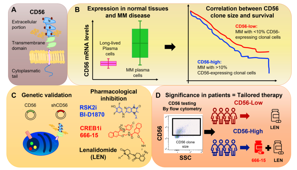
“Among others, CD56 is present at variable levels in approximately 70% of patients with multiple myeloma; however, very little is known about CD56 role in multiple myeloma.” (2022 Cottini et al.)

CD56 in MM

The role of CD56 in multiple myeloma is a topic of ongoing research and discussion among scientists and medical professionals. CD56 is a protein that is found on the surface of many different cell types, including plasma cells. Researchers have demonstrated that it plays a key role in the development and progression of multiple myeloma, making it a potential target for new treatments.

One of the main functions of CD56 is to regulate the growth and survival of plasma cells. In normal cells, CD56 helps to prevent uncontrolled growth and division of cells. However, in multiple myeloma cells, CD56 appears to play a different role. Research has shown that CD56 is overexpressed in multiple myeloma cells, leading to an increase in cell growth and division.

Additionally, CD56 has been found to play a role in the immune system’s response to cancer cells. In multiple myeloma, CD56 can suppress the immune system’s response to the abnormal cells, allowing them to continue growing unchecked. This is why multiple myeloma is often resistant to traditional cancer treatments such as chemotherapy and radiation.

Targeting CD56 in MM

There are currently several strategies being explored to target CD56 in multiple myeloma. One approach is to use drugs that block the function of CD56, in order to prevent it from promoting cell growth and division. Another approach is to use immunotherapies that stimulate the immune system to attack the abnormal cells. This can help to overcome the suppression of the immune system by CD56 and lead to more effective cancer treatment.

There is also research being conducted into the use of CAR T-cell therapy. CAR T-cell therapy involves genetically modifying a patient’s own immune cells to attack the cancer cells. In this type of therapy, the immune cells are modified to target CD56 specifically, which allows them to attack and destroy the multiple myeloma cells more effectively.

Cottini et al.

In this editorial, the researchers discuss their study, which looked at CD56-expressing clonal MM cells in more than 700 patients at the time of MM diagnosis. The researchers found that the size of these cells varied between patients and increased as the disease worsened. Results demonstrated that having a large amount of these cells was linked to worse outcomes and shorter responses to treatment. 

The study then looked at how changing the expression of CD56 affected the behavior of MM cells and found that it influenced cell growth and survival. They also discovered that a protein called RSK2 and another called CREB1 play a role in this process. They then tested medicines to block these proteins and found that they were effective in killing MM cells that had a high amount of CD56, but not as much in those with low levels of CD56.

“The authors’ preclinical data support the use of synthetic lethal approaches by CREB1/RSK2 inhibition in combination with lenalidomide, as a strategy to overcome CRBN downregulation in CD56-high MM.”

Conclusion

“In summary, this study provides a detailed description of CD56 role in MM, opening new clinically relevant scenarios.”

The role of CD56 in multiple myeloma is complex and still not fully understood. However, the researchers who wrote this editorial aimed to clearly define CD56’s key role in the development and progression of this disease, making it a potential target for new treatments. By better understanding the function of CD56, scientists and medical professionals can continue to develop new and innovative therapies to improve the lives of those affected by multiple myeloma.

“Since the majority of clinical laboratories have the capability to perform CD56 staining and define a threshold of positivity, CD56 expression can be both a prognostic and predictive factor of response to therapies, an unmet need in the MM field (Figure 1).”

Click here to read the full editorial published in Oncotarget

ONCOTARGET VIDEOS: YouTube | LabTube | Oncotarget.com

Oncotarget is an open-access, peer-reviewed journal that has published primarily oncology-focused research papers since 2010. These papers are available to readers (at no cost and free of subscription barriers) in a continuous publishing format at Oncotarget.com. Oncotarget is indexed/archived on MEDLINE / PMC / PubMed.

For media inquiries, please contact media@impactjournals.com.

Researchers Identify New Drug Candidates to Treat DLST+ Tumors

The increased expression of DLST has recently been associated with increased tumor aggression and a poor prognosis in neuroblastoma and triple-negative breast cancer.

Figure 3: Analysis of DLST-activated cell lines revealed sensitivity to protein kinase inhibiting the ERK/MAPK pathway.

The Trending With Impact series highlights Oncotarget publications attracting higher visibility among readers around the world online, in the news, and on social media—beyond normal readership levels. Look for future science news about the latest trending publications here, and at Oncotarget.com.

Listen to an audio version of this article

Dihydrolipoamide S-succinyltransferase (DLST) is a crucial gene/protein/enzyme involved in the oxidative phosphorylation (OXPHOS) pathway and cellular energy production. Recent studies have demonstrated that, in neuroblastoma and triple-negative breast cancer (TNBC), increased expression of DLST is associated with increased tumor aggression and a poor disease prognosis. Researchers also found that, in leukemia and TNBC cell lines, the knockdown of DLST leads to apoptosis. These findings suggest that neuroblastoma and TNBC may benefit from DLST-inhibiting cancer therapy.

In light of this evidence, researchers Christina Kuhn, Myriam Boeschen, Manuel Philip, Torsten Schöneberg, Doreen Thor, and Susanne Horn from the University of Leipzig, University Duisburg-Essen and the German Cancer Consortium investigated approved drugs that may target DLST-activated tumors. In their recent study, the team used data from the Genomics of Drug Sensitivity in Cancer (GDSC) project to identify new drug candidates for the treatment of DLST-activated tumors. On January 12, 2023, their research paper was published in Oncotarget’s Volume 14, entitled, “Candidate drugs associated with sensitivity of cancer cell lines with DLST amplification or high mRNA levels.”

The Study

“With the advent of complex genetic datasets of roughly 1000 cell lines in the Cancer Cell Line Encyclopedia (CCLE) and on drug resistance in the Genomics of Drug Sensitivity in Cancer project (GDSC), analyses of drug sensitivity have become possible on a larger scale [6, 7].”

Since neuroblastoma and TNBC tumor cell viability may be DLST-dependent, DLST is a promising target for cancer therapy. The researchers used the Cancer Cell Line Encyclopedia (CCLE) to identify cancer cell lines with DLST amplifications or high mRNA levels. They then measured the sensitivity of these DLST+ cell lines to 250 drugs in the GDSC dataset and compared the data to a subset of cell lines without DLST amplifications or high mRNA levels.

“To identify drugs that inhibit viability of specifically DLST-activated tumor cells, we compared cell lines with supposedly activating changes of DLST (DNA amplification, high mRNA levels) to cell lines without DLST changes.”

Results & Conclusions

“DLST-altered cell lines were more sensitive to 7 approved drugs, among these obatoclax mesylate, a BCL2 inhibitor that reduces OXPHOS in human leukemia stem cells.”

The researchers identified seven drug candidates that demonstrated significantly higher sensitivity in DLST+ cell lines than in the control cell lines. In addition to a BCL2 inhibitor found to reduce OXPHOS, multiple protein kinase inhibitors were identified as efficient in the DLST+ cell lines. This suggests that DLST-altered cell lines may also be vulnerable to ERK/MAPK pathway-targeting drugs. The researchers propose that the drug candidates identified in this study warrant further drug efficacy testing in knock-in cell lines and DLST-activated tumors.

“We therefore conclude that, in addition to OXPHOS, protein kinases could be potential targets of therapy in the presence of DLST amplifications or high mRNA levels.”

Click here to read the full research paper published by Oncotarget

ONCOTARGET VIDEOS: YouTube | LabTube | Oncotarget.com

Oncotarget is an open-access, peer reviewed journal that has published primarily oncology-focused research papers since 2010. These papers are available to readers (at no cost and free of subscription barriers) in a continuous publishing format at Oncotarget.com. Oncotarget is indexed/archived on MEDLINE / PMC / PubMed.

For media inquiries, please contact media@impactjournals.com.

Novel Antibody Drug Conjugate Improves Murine Acute Myeloid Leukemia

Researchers from Astellas Pharma Inc. investigated the efficacy of a novel antibody drug conjugate combined with venetoclax and azacitidine in a mouse model of acute myeloid leukemia.

Novel Antibody Drug Conjugate Improves Murine Acute Myeloid Leukemia

Listen to an audio version of this article

The average age of patients with acute myeloid leukemia (AML) is 67 years old. Older adults generally have a lower tolerance for treatments that exhibit high off-target toxicity. Additionally, chemotherapy-relapsed or -refractory (R/R) AML patients are often at an advanced stage of disease and are therefore more likely to have comorbidities that may reduce their tolerance for harsh treatments.

Thus, pharmaceutical AML drugs with high efficacy and low toxicity are in high demand. Antibody drug conjugates (ADCs) are emerging as promising therapeutic approaches to more safely treat hematological malignancies by reducing side effects. ADCs are designed to decrease damage to healthy tissues by specifically targeting tumor-associated antigens attached to cancer cells.

“Antibody drug conjugates (ADC) are one of the modalities that aims to dissociate drug efficacy from toxicity. ADC consists of three components: antibody specific for tumor associated antigen, drug linker and cytotoxic payload.”

Astellas Pharma

Recently, researchers from Astellas Pharma Inc. (a pharmaceutical company in Japan) developed ASP1235—a novel ADC that targets Fms-like tyrosine kinase 3 (FLT3). In more than 90% of AML patients, FLT3 is overexpressed on leukemic blasts. ASP1235 is designed to target FLT3-positive leukemia cells and deliver the cytotoxic drug payload to these cells. However, this drug alone was found to have only a mild effect on AML cells, prompting researchers to assess the efficacy of ASP1235 in combination with other drugs.

In a new study, Astellas Pharma researchers Hirofumi Tsuzuki, Tatsuya Kawase, Taisuke Nakazawa, Masamichi Mori, and Taku Yoshida investigated the efficacy of ASP1235 combined with venetoclax (an anti-apoptotic agent) and azacitidine (a DNA methyltransferase inhibitor) in an experimental mouse model of AML. Their research paper was published in Oncotarget on December 20, 2022, and entitled, “Anti-tumor effect of antibody drug conjugate ASP1235 targeting Fms-like tyrosine kinase 3 with venetoclax plus azacitidine in an acute myeloid leukemia xenograft mouse model.”

“In this study, we sought to evaluate the therapeutic effect of ASP1235 in combination with venetoclax plus azacitidine, a novel standard-of-care treatment for elderly AML patients, in ASP1235 poor sensitive AML cells.”

The Study

The researchers first aimed to determine an AML cell line that was only partially sensitive to ASP1235 monotherapy. They determined the THP-1 cell line was appropriate for further investigation. They compared FLT3 and Bcl-2 expression levels in THP-1 cells with primary leukemic cells from chemotherapy R/R AML patients to consider the clinical relevance of each. In THP-1 cells, the expression levels of FLT3 and Bcl-2 were found to be clinically relevant.

“It has been reported that the proportion of patients showing high Bcl-2 expression was greater in chemotherapy R/R AML patients compared to that in newly diagnosed patients [4]. Thus, we investigated the expression levels of Bcl-2 together with FLT3 to further consider the relevance of THP-1 cells for evaluation on the combination treatment with venetoclax.”

To confirm their in vitro findings, they used a THP-1 xenograft mouse model for in vivo investigation of ASP1235 sensitivity. Their findings indicated that the THP-1 cell was a partially sensitive preclinical model to ASP1235. Next, the researchers evaluated the in vivo efficacy of ASP1235 in combination with venetoclax plus azacitidine using the THP-1 xenograft mouse model. The results showed that the combination therapy induced a significant reduction in tumor size compared to ASP1235 monotherapy and the other two drugs alone. This suggests that ASP1235 has an enhanced anti-tumor effect in combination with venetoclax and azacitidine.

“Consistent with in vitro observations in Figure 4, triple combination treatment with ASP1235, venetoclax and azacitidine induced tumor regression, and the anti-tumor effect of the triple combination was much stronger than that of ASP1235 single agent or venetoclax plus azacitidine without obvious body weight loss (Figure 5).”

Figure 5: ASP1235 showed enhanced anti-tumor effect in combination with venetoclax and azacitidine in THP-1 xenograft mouse model.

Conclusions

The findings of this study suggest that the combination therapy of ASP1235, venetoclax and azacitidine can be an effective treatment option for elderly patients or patients with chemotherapy R/R AML. This combination therapy induced a significant reduction in xenograft tumors in the THP-1 mouse model, suggesting that it may be a promising therapeutic approach for AML patients. Further clinical trials are needed to confirm these results.

“In conclusion, the triple combination treatment of ASP1235, venetoclax and azacitidine has the potential to benefit AML patients, and there is a possibility to expect the combination effect of ASP1235 and venetoclax regimen in FLT3 positive cancers beyond AML.”

Click here to read the full research paper published by Oncotarget

ONCOTARGET VIDEOS: YouTube | LabTube | Oncotarget.com

Oncotarget is an open-access journal that publishes primarily oncology-focused research papers in a continuous publishing format. These papers are available at no cost to readers on Oncotarget.com. Open-access journals have the power to benefit humanity from the inside out by rapidly disseminating information that may be freely shared with researchers, colleagues, family, and friends around the world.

For media inquiries, please contact media@impactjournals.com.

A New Method of Targeting Exosomes in Precision Medicine

Oncotarget published a new editorial perspective by Dr. Mujib Ullah, entitled, “The future of bioorthogonal-chemistry for targeting of exosomes in precision medicine.”

A New Method of Targeting Exosomes in Precision Medicine
Figure 1: Schematic illustration showing tracking of exosomes labeled by phospholipid-based bioorthogonal conjugation.
Listen to an audio version of this article

Extracellular vesicles are membrane-bound vehicles released by cells into the extracellular environment. There are three known types of extracellular vesicles: microvesicles, apoptotic bodies and exosomes. Discovered in 1983, exosomes can be defined as packets of bio-nanoparticles released by cells containing bioactive molecules such as proteins, lipids and nucleic acids. Exosomes can deliver their payload to other cells and are now also recognized for their role in cell-to-cell communication. This makes exosomes attractive targets for precision medicine tactics. However, targeting exosomes is challenging due to their nano-size and reactive contents. Bioorthogonal-chemistry may provide a new approach for targeting exosomes in precision medicine.

“Bioorthogonal is the name of a chemical reaction that can occur inside of living cells without interfering the naïve biological process [1, 2].”

Bioorthogonal-chemistry allows for the attachment of bioactive molecules to the surface of exosomes without disturbing the native environment. Developed in the early 2000s, this strategy could potentially be used to deliver therapeutic drugs or bioactive molecules directly to the target site with high precision. Bioorthogonal-chemistry is still at an early stage of development, but it holds promise in precision medicine for the treatment of cancer and other illnesses. By providing a way to target exosomes with bioactive molecules, bioorthogonal-chemistry could help to significantly improve the efficacy of medical treatments. It could also reduce the side effects of current treatments and increase safety for patients.

“The concept of bioorthogonal chemistry has inspired a generation of biologists to think about RNA editing and bioengineering of exosomes [3, 4].”

On December 6, 2022, Oncotarget published a new editorial perspective by Dr. Mujib Ullah from Stanford University, entitled, “The future of bioorthogonal-chemistry for targeting of exosomes in precision medicine.”

In this short editorial perspective, Dr. Ullah discussed current exosome-loading techniques, including electroporation, heat shock, sonication, and ultracentrifugation. He wrote that these techniques are disruptive and potentially ineffective methods of exosome loading. On the other hand, he explained that using a precise, targeted bioorthogonal reaction can overcome the aforementioned issues. Dr. Ullah listed nine questions at the end of his editorial perspective. He believes these important questions must be answered in order to showcase the potential of bioorthogonal reactions in future clinical biomedical applications:

  1. Can bioorthogonal chemistry help in the development of more powerful bioimaging and biosensing techniques?
  2. Can the combination of exosomes with biorthogonal chemistry overcome some of the current translational hurdles in precision medicine?
  3. Can new drugs be designed inside humans?
  4. Can the exosomes cargo be detected by conjugation chemistry?
  5. Can we track the metabolites encapsulated inside the exosomes?
  6. Can pharmaceuticals be synthesized inside living system?
  7. How many orthogonal reactions can be performed in one time?
  8. What is the half life of these reactions?
  9. Biorthogonal reactions are rapid but what is the speed of reactions? 

Click here to read the full editorial perspective published by Oncotarget

ONCOTARGET VIDEOS: YouTube | LabTube | Oncotarget.com

Oncotarget is an open-access journal that publishes primarily oncology-focused research papers in a continuous publishing format. These papers are available at no cost to readers on Oncotarget.com. Open-access journals have the power to benefit humanity from the inside out by rapidly disseminating information that may be freely shared with researchers, colleagues, family, and friends around the world.

For media inquiries, please contact media@impactjournals.com.

Plasma Growth Hormone in HCC: A Biomarker of Response to Atezo/Bev?

In a new study, researchers investigated the plasma growth hormone as a potential biomarker of response to atezolizumab and bevacizumab in advanced hepatocellular carcinoma patients.

Plasma Growth Hormone in HCC: A Biomarker of Response to Atezo/Bev?

The Trending With Impact series highlights Oncotarget publications attracting higher visibility among readers around the world online, in the news, and on social media—beyond normal readership levels. Look for future science news about the latest trending publications here, and at Oncotarget.com.

Listen to an audio version of this article

Hepatocellular carcinoma (HCC) is a highly aggressive cancer of the liver with a very poor prognosis; many patients pass away within a year of diagnosis. Currently, there is no effective screening method for HCC and thus, 80% of patients are diagnosed at advanced stages. This makes treatment difficult and often unsuccessful. As a result, new treatments for HCC are constantly being explored.

Atezolizumab and bevacizumab are two standard therapies used to treat unresectable, advanced HCC. However, researchers Yehia I. Mohamed, Dan G. Duda, Muhammad O. Awiwi, Sunyoung S. Lee, Lina Altameemi, Lianchun Xiao, Jeffrey S. Morris, Robert A. Wolff, Khaled M. Elsayes, Rikita I. Hatia, Aliya Qayyum, Shadi M. Chamseddine, Asif Rashid, James C. Yao, Armeen Mahvash, Manal M. Hassan, Hesham M. Amin, and Ahmed Omar Kaseb from MD Anderson Cancer Center, Massachusetts General Hospital, Harvard Medical School, Michigan State University, and University of Pennsylvania Perelman School of Medicine noticed a significant gap in research on biomarkers of response in advanced HCC patients treated with atezolizumab plus bevacizumab. The team conducted a new study aimed at beginning to close this gap. On December 6, 2022, their research paper was published in Oncotarget’s Volume 13, entitled, “Plasma growth hormone is a potential biomarker of response to atezolizumab and bevacizumab in advanced hepatocellular carcinoma patients.”

“This study investigated the biomarker value of plasma growth hormone (GH) level as a potential biomarker to predict outcome in unresectable HCC patients treated with current standard therapy, atezolizumab plus bevacizumab (Atezo/Bev).”

The Study

Plasma growth hormone (GH) is a potential biomarker that had not previously been evaluated in relation to this treatment regimen in HCC before. In this study, the researchers included 37 patients with advanced HCC. The patients received atezolizumab plus bevacizumab at the MD Anderson Cancer Center between June 2018 and November 2021. The median age of the patients was 67 years old, and the vast majority were male (83.8%). 

The team measured plasma GH levels, progression-free survival (PFS) and overall survival (OS). Plasma GH levels were measured by ELISA and used to stratify the HCC patients into GH-high and GH-low groups. The Kaplan-Meier method was used to calculate median OS and PFS. The Log-rank test was used to compare survival outcomes between GH-high and -low groups.

“To the best of our knowledge, this is the first study to assess clinical prognostic value of plasma GH level in patients who have received atezolizumab plus bevacizumab in clinical setting.”

The results of the study showed that plasma GH levels significantly correlated with OS. At the time of the analysis, the one-year survival rate was 70% among GH-low patients and 33% among GH-high patients. OS was significantly superior in GH-low compared to GH-high patients. PFS showed a non-significant trend in favor of GH-low patients compared to the GH-high group.

Conclusion

“Despite the small data size, plasma GH levels were strongly predictive of the disease outcome in patients treated with Atezo/Bev.”

The study concluded that plasma GH levels may be a promising biomarker for predicting response to atezolizumab plus bevacizumab in advanced HCC patients. Further, plasma GH levels may be used to stratify advanced HCC patients into high- and low-risk groups. The researchers recommend further research in larger scale and different populations to validate the findings and explore plasma GH levels as a potential biomarker of response to this treatment regimen in HCC.

“In conclusion, our study demonstrate[s] that plasma GH represents a candidate biomarker for predicting treatment outcomes in patients with advanced HCC treated with Atezo/Bev. Future studies in larger randomized clinical trial and with a more diverse ethnic, race, and gender background are warranted to further validate these findings.”

Click here to read the full research paper published by Oncotarget

ONCOTARGET VIDEOS: YouTube | LabTube | Oncotarget.com

Oncotarget is an open-access journal that publishes primarily oncology-focused research papers in a continuous publishing format. These papers are available at no cost to readers on Oncotarget.com. Open-access journals have the power to benefit humanity from the inside out by rapidly disseminating information that may be freely shared with researchers, colleagues, family, and friends around the world.

For media inquiries, please contact media@impactjournals.com.

Biomarkers May Predict Neoadjuvant Chemosensitivity in Bladder Cancer

In a new study, researchers aimed to identify and validate predictive biomarkers of response to neoadjuvant chemotherapy (NAC) in patients with muscle-invasive bladder cancer (MIBC).

Biomarkers May Predict Neoadjuvant Chemosensitivity in Bladder Cancer
Listen to an audio version of this article

Neoadjuvant chemotherapy (NAC) is a type of cancer treatment involving the administration of chemotherapy drugs before surgery. The goal of NAC is to shrink the tumor(s) in order to make it/them easier to remove during surgery and to decrease the chance of cancer recurrence after treatment. NAC is typically well tolerated by patients and has been shown to improve outcomes in patients with bladder cancer.

Predictive biomarkers are being increasingly used in oncology to identify patients who are likely to respond to chemotherapy. In the past, the decision to administer chemotherapy was based on tumor type and stage. However, it is now understood that there is considerable heterogeneity within these groups, and that not all patients will respond to the same treatment. Predictive biomarkers can help to overcome this challenge by identifying those patients who are most likely to benefit from chemotherapy.

There are a number of different types of predictive biomarkers, which can be divided into two broad categories: tumor biomarkers and host biomarkers. Tumor biomarkers are usually specific to the tumor type and can include markers of cell proliferation and DNA repair. Host biomarkers are usually found in the blood or other bodily fluids and can include markers of inflammation, immune function and metabolism. The use of predictive biomarkers has the potential to improve the efficacy of chemotherapy and reduce toxicity by avoiding its use in patients who are unlikely to benefit.

The Study

In a new study, researchers Neal Murphy, Andrew J. Shih, Paras Shah, Oksana Yaskiv, Houman Khalili, Anthony Liew, Annette T. Lee, and Xin-Hua Zhu from Donald and Barbara Zucker School of Medicine at Hofstra/Northwell, Northwell Health Cancer Institute, Feinstein Institutes for Medical Research, and Mayo Clinic aimed to develop and validate a predictive biomarker panel for response to NAC in patients with muscle-invasive bladder cancer (MIBC). Their research paper was published on November 2, 2022, in Oncotarget’s Volume 13, entitled, “Predictive molecular biomarkers for determining neoadjuvant chemosensitivity in muscle invasive bladder cancer.”

“The NAC non-responders suffer from unnecessary adverse effects and a delay in time to cystectomy leading to worse overall survival [9, 10]. Subsequently, there remains a critical need to understand the molecular biology behind NAC responsiveness, in order to better tailor individual NAC therapy.”

The purpose of this research was to “develop a molecular signature that can identify MIBC NAC responders (R) and non-responders (NR) using a cohort of known NAC response phenotypes, and better understand differences in molecular pathways and subtype classifications between NAC R and NR.” Researchers identified a total of 26 patients with known NAC response for inclusion in this study. These patients were assigned at random to either the discovery or validation cohort. The discovery cohort consisted of seven NAC responders and 11 non-responders. The validation cohort consisted of three responders and five non-responders.

Transurethral resection of bladder tumor (TURBT) specimens from the Northwell Health pathology department were received as formalin-fixed, paraffin-embedded (FFPE) tissue blocks. Pathologic response was determined at the time of cystectomy. Messenger RNA (mRNA) and microRNA (miRNA) from the FFPE blocks were sequenced using RNAseq and qPCR, respectively.

“To our knowledge, our study is the first to use combined differential mRNA and miRNA expression in MIBC to identify a NAC response signature.”

The Results

“We report significant gene sets associated with NAC response phenotype, as well as three multigene and miRNA signatures generated by CCA that can be used to potentially classify NAC response.”

In the discovery cohort, the researchers found that 2309 genes were differentially expressed between the NAC responders and non-responders. In the validation cohort, 602 genes and 13 miRNA were differentially expressed. Canonical correlation (CC) analysis found that three CCs (CC13: nucleoside triphosphate metabolic process; CC16: cell cycle and cellular response to DNA damage; and CC17: DNA packaging complex) were differentiated in the discovery and validation datasets. As far as MIBC subtypes, the MD Anderson p53-like subtype, CIT MC4 subtype and Consensus Class stroma-rich subtype had the strongest correlation with a non-responder phenotype. There were no subtypes that had strong correlations with the responder phenotype.

“In conclusion, our results identify molecular signatures that can be used to differentiate MIBC NAC responders versus non-responders. We have presented the salient molecular pathways and relevant genes, including mitochondrial response gene expression (MRPS12, MRPS34, MRPS28, MRPS14, and MRPS2), DNA replication initiation, and DNA unwinding and DNA damage (MCM2-3, MCM5-6 and XAP , ELK4, and FOXA3) that can be further analyzed to better understand NAC response. The above mentioned genes derived from their respective three pathways may be selected as part of a NAC response biomarker panel. In addition, we have highlighted the utility of molecular subtyping in relation to NAC response. If validated in a larger cohort, these findings may help deliver chemotherapy to those patients most likely to respond.”

Conclusion

Neoadjuvant chemotherapy is a promising treatment option for muscle-invasive bladder cancer patients, however, there is a class of patients who do not respond to chemotherapy. The results of this study implicate several different types of biomarkers that may be associated with chemosensitivity in MIBC patients. Further research is needed to validate these findings. Ultimately, if validated, these biomarkers could help to spare non-responders from side effects associated with ineffective and unnecessary chemotherapy.

“Our results identify molecular signatures that can be used to differentiate MIBC NAC R versus NR, salient molecular pathway differences, and highlight the utility of molecular subtyping in relation to NAC response.”

Click here to read the full research paper published by Oncotarget

ONCOTARGET VIDEOS: YouTube | LabTube | Oncotarget.com

Oncotarget is an open-access journal that publishes primarily oncology-focused research papers in a continuous publishing format. These papers are available at no cost to readers on Oncotarget.com. Open-access journals have the power to benefit humanity from the inside out by rapidly disseminating information that may be freely shared with researchers, colleagues, family, and friends around the world.

For media inquiries, please contact media@impactjournals.com.

New Target Fights Ferroptosis- and Radio-Resistance in Lung Cancers

Researchers published a research perspective about a recent study that uncovered FSP1 as a novel target gene that mediates ferroptosis resistance and radioresistance in lung cancer cells.

Figure 1: KEAP1-NRF2 axis and its molecular effectors in the regulation of ferroptosis.
Listen to an audio version of this article

Ferroptosis is a type of cell death caused by the accumulation of iron and lipid peroxides in cells. Cancer cells are often resistant to ferroptosis, which allows them to survive and proliferate. Radioresistance is another common feature of cancer cells that allows them to resist the effects of radiation therapy.

A new research paper (published on April 22, 2022) identified ferroptosis suppressor protein 1 (FSP1) as a novel KEAP1/NRF2 target gene and demonstrated that FSP1 plays an essential role in NRF2-mediated ferroptosis resistance and radioresistance in KEAP1-deficient lung cancer cells. 

Recently, researchers Nsengiyumva Emmanuel, Hongen Li, Jing Chen, and Yilei Zhang from Xi’an Jiaotong University, Ruyang People’s Hospital and Shaanxi Jiuzhou Biomedical Science and Technology Group wrote a paper about the implications of these findings. On October 19, 2022, their research perspective was published in Oncotarget’s Volume 13, entitled, “FSP1, a novel KEAP1/NRF2 target gene regulating ferroptosis and radioresistance in lung cancers.”

“In a recent study by Pranavi Koppula et al. from The University of Texas MD Anderson Cancer Center, FSP1 was demonstrated as a novel target of NRF2 and to play a vital role in KEAP1/NRF2-mediated ferroptosis regulation [13], which reveals the important role of genetic regulation of FSP1 in cancer development.”

The KEAP1-NRF2 regulatory pathway is crucial for protecting cells against oxidative damage. In lung cancer cells, KEAP1 mutations lead to NRF2 deregulation and contribute to tumorigenesis. Researchers have been searching for an effective way of targeting this pathway in cancer cells to sensitize them to ferroptosis/radiation-inducing agents. They found that FSP1 is upregulated in KEAP1-mutant lung cancer cells, and its expression is associated with resistance to ferroptosis and radiotherapy. Coenzyme Q10- (CoQ) FSP1 signaling represents a potential target for overcoming resistance to ferroptosis and radiotherapy in KEAP1-mutant lung cancer cells.

“Currently, there are four major ferroptosis-defending systems, while NRF2 could directly control two of them, SLC7A11/GSH/GPX4 axis and CoQ/FSP1 axis.”

This study provides new insights into the role of target genes in the KEAP1-NRF2 pathway and how they contribute to cancer cells’ resistance to ferroptosis and radiotherapy. However, the mechanism by which KEAP1-NRF2 target genes mediate resistance to ferroptosis is not yet fully understood. Fortunately, the identification of FSP1 as a key target gene involved in this process opens up new therapeutic avenues for the treatment of lung cancer.

“Here, Pranavi Koppula and her colleagues’ study indicates that pharmacological targeting of CoQ-FSP1 signaling to overcome KEAP1 deficiency-induced radioresistance could be a potentially effective therapeutic strategy in treating KEAP1 mutant lung cancers.”

Click here to read the full research perspective published by Oncotarget

ONCOTARGET VIDEOS: YouTube | LabTube | Oncotarget.com

Oncotarget is an open-access journal that publishes primarily oncology-focused research papers in a continuous publishing format. These papers are available at no cost to readers on Oncotarget.com. Open-access journals have the power to benefit humanity from the inside out by rapidly disseminating information that may be freely shared with researchers, colleagues, family, and friends around the world.

For media inquiries, please contact media@impactjournals.com.

Synergy of HDACi, PARPi and Chemotherapeutics Against Blood Cancer

Researchers investigated the efficacy of HDAC inhibitors in combination with PARP inhibitors and chemotherapeutic drugs in multiple blood cancer cell lines.

Synergy of HDACi, PARPi and Chemotherapeutics Against Blood Cancer
Listen to an audio version of this article

Chromatin constitutes chromosomes in eukaryotic cells and comprises DNA and proteins. Chromosomes produce proteins and enzymes that are essential for cellular function and maintenance, including DNA repair. A critical process for DNA repair is poly(ADP-ribosyl)ation, or PARylation.

PARylation is triggered by poly(ADP ribose) polymerase (PARP) enzymes. When DNA becomes damaged, PARP enzymes bind to the damaged location in the cell. In cancer cells, however, this natural process can be counterproductive in respect to cancer treatment. PARylation can produce DNA repair mechanisms in cancer cells that can lead to cell death evasion and even drug resistance. Inhibiting PARylation may be a viable therapeutic strategy for cancer treatment.

HDAC Inhibitors

Histones, the main proteins that constitute chromatin, undergo post-translational modifications that regulate gene expression. Histone acetylation is an important epigenetic process that affects gene expression by relaxing the chromatin structure, making chromatin remodeling more feasible. Histone deacetylases (HDACs) are enzymes that can have the opposite effect. Histone deacetylation makes the chromatin more compact and difficult to remodel. The overexpression of HDAC has also been associated with tumorigenesis. Histone deacetylase inhibitors (HDACi) are a class of therapeutics that have shown promise in the treatment of hematologic malignancies (blood cancer) and solid tumors.

“Overexpression of HDACs has been associated with tumorigenesis by down-regulation of tumor suppressor genes [3, 4]; hence, HDAC inhibitors (HDACi) including vorinostat (SAHA), romidepsin (Rom), panobinostat (Pano) and belinostat have been approved by the United States Food and Drug Administration for the treatment of hematologic and other malignancies [5]. These inhibitors restore appropriate gene expression, resulting in induction of cell differentiation, cell cycle arrest and apoptosis [6].”

The Study

In a new study, researchers Benigno C. Valdez, Yago Nieto, Bin Yuan, David Murray, and Borje S. Andersson from the Department of Stem Cell Transplantation and Cellular Therapy at the University of Texas MD Anderson Cancer Center and the Cross Cancer Institute’s Department of Experimental Oncology at the University of Alberta investigate the efficacy of HDACi in combination with PARP inhibitors (PARPi) and chemotherapeutic drugs to treat hematologic cancer. On October 14, 2022, their research paper was published in Volume 13 of Oncotarget, entitled, “HDAC inhibitors suppress protein poly(ADP-ribosyl)ation and DNA repair protein levels and phosphorylation status in hematologic cancer cells: implications for their use in combination with PARP inhibitors and chemotherapeutic drugs.”

“Despite their preclinical efficacy, HDACi do not seem to be clinically highly effective as monotherapy, and potentially more effective anti-tumor activity is observed when they are combined with other anti-cancer drugs [79].”

Studies on the interactions of HDACi with PARPi in cancers of the blood are limited, especially when combined with chemotherapeutic agents. The researchers used a panel of hematologic cancer cell lines (acute myeloid leukemia, T-cell acute lymphoblastic leukemia, chronic myeloid leukemia, and multiple myeloma) and patient-derived cell samples to study the effect of HDACi (including SAHA (Vorinostat), panobinostat (Pano), romidepsin (Rom) and trichostatin A (TSA)) on PARylation. In addition, the team looked at the efficacy of HDACi combined with PARPi, including Olaparib (Ola) and niraparib (Npb), and with chemotherapeutic agents gemcitabine (Gem), busulfan (Bu) and melphalan (Mel).

Results

The researchers found that hematologic cancer cell lines and patient-derived cell samples exposed to various HDACi resulted in a significant caspase-independent inhibition of protein PARylation. HDACi-mediated inhibition of protein PARylation was mainly catalyzed by PARP1. These findings suggest that HDACi could potentially be used in combination with PARP inhibitors and chemotherapeutic drugs to treat blood cancers.

“Our results indicate that the anti-tumor efficacy of HDACi is partly due to down-regulation of PARylation, which negatively affects the status of DNA repair proteins. This repair inhibition, combined with the high levels of oxidative and DNA replication stress characteristic of cancer cells, could have conferred these hematologic cancer cells not only with a high sensitivity to HDACi but also with a heightened dependence on PARP and therefore with extreme sensitivity to combined HDACi/PARPi treatment and, by extension, to their combination with conventional DNA-damaging chemotherapeutic agents. The observed synergism of these drugs could have a major significance in improving treatment of these cancers.”

Conclusion

HDACi drugs can inhibit PARylation. The combination of HDACi-mediated inhibition of PARylation was complemented by PARPi and chemotherapeutic agents in multiple blood cancer cell lines. The efficacy of this combined treatment was superior to that of any single agent, supporting the further clinical development of HDACi in cancer therapy. These findings could potentially be used to improve the treatment of hematologic cancers.

“In conclusion, our results provide a molecular explanation for the HDACi-mediated inhibition of DNA repair in hematologic cancer cells and support the combinatorial application of HDACi, PARPi and chemotherapeutic agents for the treatment of hematologic malignancies.”

Click here to read the full research paper published by Oncotarget

ONCOTARGET VIDEOS: YouTube | LabTube | Oncotarget.com

Oncotarget is an open-access journal that publishes primarily oncology-focused research papers in a continuous publishing format. These papers are available at no cost to readers on Oncotarget.com. Open-access journals have the power to benefit humanity from the inside out by rapidly disseminating information that may be freely shared with researchers, colleagues, family, and friends around the world.

For media inquiries, please contact media@impactjournals.com.

CRISPR Screens Identify Novel Targets for Personalized Cancer Therapy

In this trending new study, researchers used CRISPR-based genome-wide screens to identify genetic determinants of PARP10-mediated cellular survival.

CRISPR Screens Identify Novel Targets for Personalized Cancer Therapy
Listen to an audio version of this article

The Trending With Impact series highlights Oncotarget publications attracting higher visibility among readers around the world online, in the news, and on social media—beyond normal readership levels. Look for future science news about the latest trending publications here, and at Oncotarget.com.

Genetic interactions involved in the survival of cancer cells are potential therapeutic targets in personalized cancer therapy. “Synthetic lethal” is a type of genetic interaction where the knockout of one gene can cause cell death but only in the presence of another dependent gene. Cancer researchers view synthetic lethality screening as a powerful tool in precision medicine.

“Identifying genetic susceptibilities based on PARP10 expression levels is thus potentially relevant for finding new targets for precision oncology.”

Poly-ADP-ribose polymerase 10, or PARP10, is a nuclear protein that is overexpressed in multiple cancers. Genetic susceptibilities based on PARP10 expression levels in an individual may be potential targets for personalized cancer therapy. In a new study, researchers Jude B. Khatib, Emily M. Schleicher, Lindsey M. Jackson, Ashna Dhoonmoon, George-Lucian Moldovan, and Claudia M. Nicolae, from the Department of Biochemistry and Molecular Biology at Penn State College of Medicine, used CRISPR-based, genome-wide genetic screens to identify potential synthetic lethality interactions with PARP10-overexpressing and -knockout cancer cells. On September 28, 2022, their research paper was published in Oncotarget and entitled, “Complementary CRISPR genome-wide genetic screens in PARP10-knockout and overexpressing cells identify synthetic interactions for PARP10-mediated cellular survival.”

“Here, we employed complementary CRISPR loss-of-function genome-wide screening to identify genes required for proliferation of PARP10-overexpressing and PARP10-knockout cells.”

The Study

To identify potential synthetic lethal targets, the researchers conducted a CRISPR-based, genome-wide genetic screen of both PARP10-overexpressing and PARP10-knockout tumorigenic and non-tumorigenic breast cells. The screen looked for genes that were required for cell proliferation in the presence of PARP10 overexpression or PARP10 knockout.

“Here, we performed a series of CRISPR genome-wide loss-of-function screens in isogenic control and PARP10-overexpressing or PARP10-knockout cell lines, to identify genetic determinants of PARP10-mediated cellular survival.”

In the PARP10 overexpressing cells, the top results from their CRISPR screen were validated with biological pathway enrichment analyses, using both KEGG and Gene Ontology databases. A functional interaction between ATM and PARP10 expression was found. ATM promoted cell proliferation in PARP10-overexpressing cells.

In the genome-wide CRISPR knockout screens, genes required for the viability of PARP10-knockout cells were identified. In the PARP10 knockout cells, the top results from their CRISPR screen were validated with biological pathway enrichment analyses, using both KEGG and Gene Ontology databases. They identified the CDK2-Cyclin E1 complex as a genetic determinant for the proliferation of PARP10-knockout cells.

“Our work identifies a network of functionally relevant PARP10 synthetic interactions, and reveals a set of factors which can potentially be targeted in personalized cancer therapy.”

Conclusion

The researchers identified several genes that were differentially required for cell proliferation in the presence of PARP10 overexpression or knockout. Some of these genes have been previously implicated in cancer, while others were novel candidate cancer targets. The identification of these potential synthetic lethal interactions provides new insights into the role of PARP10 in cancer and may be useful for precision oncology. This study highlights the importance of using complementary CRISPR-based screens to identify potential cancer targets.

“We found that DNA repair factors, including ATM, a master regulator of the DNA damage checkpoint response, are specifically promoting the proliferation of PARP10-overexpressing cells. Moreover, we identified a role for PARP10 in regulating ATM recruitment to stressed replication forks. Finally, we found that the CDK2-cyclin E1 complex is specifically required for the proliferation of PARP10-deficient cells. Our work reveals novel PARP10 genetic interactions of functional relevance and identifies a set of factors which can potentially be targeted in personalized cancer therapy.”

Click here to read the full research paper published by Oncotarget

ONCOTARGET VIDEOS: YouTube | LabTube | Oncotarget.com

Oncotarget is an open-access journal that publishes primarily oncology-focused research papers in a continuous publishing format. These papers are available at no cost to readers on Oncotarget.com. Open-access journals have the power to benefit humanity from the inside out by rapidly disseminating information that may be freely shared with researchers, colleagues, family, and friends around the world.

For media inquiries, please contact media@impactjournals.com.

How High Altitudes Influence HIF-1, Gastric Cancer & Patient Survival

In a new study, researchers evaluated the high altitude in Ecuador and how it may influence HIF-1 expression and the survival of Ecuadorian patients with gastric cancer.

El Panecillo hill in Quito, Ecuador
Listen to an audio version of this article

Altitude is considered any elevation above sea level. Higher altitude environments are known to influence various physiological processes in the human body, including those related to hypoxia-inducing factors (HIF), vitamin D, ultraviolet radiation, oxygen toxicity, and changes in pH. Researchers have suggested that altitude may even affect the development and progression of some diseases, including stomach/gastric cancer.

“Gastric cancer is the third leading cause of death in the world and is estimated to cause almost 15 million deaths by 2035 [2].”

Gastric Cancer & Altitude

The primary subtype of gastric cancer is gastric adenocarcinoma (GA). GA develops in the mucus-secreting cells that line the stomach (gastric epithelium). Higher incidence rates of GA have been found among populations living at high altitudes. High altitudes are notorious for low air pressure and decreased oxygen saturation levels. Decreases in oxygen (hypoxia) can activate the transcriptional regulator hypoxia-inducing factor-1 (HIF-1). HIF-1 is known to be upregulated in a variety of human cancers, including GA. The role of HIF-1 in GA pathogenesis and prognosis has not yet been fully understood.

“Gastric adenocarcinoma (GA) has a high incidence in Ecuador, in men it ranks third and in women it ranks fifth.”

There is a higher incidence of GA among people living in Ecuador. This is a country that straddles the equator yet, the altitude in Ecuador varies significantly across the country. For example, the altitude is 2,850 meters in the capital city of Quito (the second-highest capital city in the world). In Guayaquil (a coastal city in Ecuador) the altitude is only nine meters. These facts make Ecuador an optimal location for studying the effects of altitude on gastric adenocarcinoma.

“Ecuador has a varied altitude diversity and there is a differential incidence of cancer between populations living in the Andean or mountainous region when compared to coastal populations or living at low altitude.”

The Study

In a new retrospective study, researchers Edwin Cevallos Barrera, Edson Zangiacomi Martinez, Mariangela Ottoboni Brunaldi, Eduardo Antonio Donadi, Ajith Kumar Sankarankutty, Rafael Kemp, and José Sebastiao dos Santos from Universidad Central del Ecuador and University of São Paulo evaluated the high altitude in Ecuador and how it may influence HIF-1 expression and the survival of Ecuadorian patients with GA. Their research paper was published on September 14, 2022, in Oncotarget’s Volume 13, entitled, “Influence of high altitude on the expression of HIF-1 and on the prognosis of Ecuadorian patients with gastric adenocarcinoma.”

A total of 229 Ecuadorians were assessed in this study. The researchers included 155 cases of GA; 99 of the individuals with GA lived in the mountainous regions of Quito and Ambato, and 56 individuals lived in the coastal region of Guayaquil. (Controls accounted for 74 people; 25 from the coast and 49 from the mountains.) The team followed-up with all individuals in this study from 2005 to 2018 and collected blood and tissue samples. They performed immunohistochemistry and other analyses to evaluate HER2 and HIF-1 expression.

“Analyses were performed using Fisher’s exact and Breslow-Day tests for homogeneity and Kaplan-Meier curves and restricted median survival time ΔRMST.”

Results & Conclusion

After 10 years, median survival was significantly higher among GA patients living along the coast. In the GA samples, HIF-1 was observed in 66.1% of the coastal patients and in 43.4% of the mountainous patients. Positive HIF-1 expression was associated with improved survival among GA patients living in the mountains. Interestingly, in the control group (without GA), HIF-1 expression was observed in 95.9% of the mountainous samples and only 32% of the coastal samples. Their results showed that 89% of the control population exhibited gastritis.

The researchers were forthcoming about the limitations of this study. Differences in quantities and the male-to-female ratios within the GA and control coastal and mountainous groups may have influenced data. Despite these limitations, the researchers observed that coastal GA patients and individuals who expressed HIF-1 had a better prognosis. However, HIF-1 expression was only associated with better survival in the mountain region. These findings suggest that HIF-1 expression may be a protective factor against GA progression in people living at high altitudes.

“Concluding, this study suggests that HIF-1 has a differential expression pattern in gastric samples according to geographical features, being highly expressed even in non-carcinomatous cells (gastritis and normal mucosa) from individuals living in regions of high altitude, indicating that the gastric HIF-1 expression may be an adaptation of the individual to high altitudes.”

Click here to read the full research paper published by Oncotarget

ONCOTARGET VIDEOS: YouTube | LabTube | Oncotarget.com

Oncotarget is an open-access journal that publishes primarily oncology-focused research papers in a continuous publishing format. These papers are available at no cost to readers on Oncotarget.com. Open-access journals have the power to benefit humanity from the inside out by rapidly disseminating information that may be freely shared with researchers, colleagues, family, and friends around the world.

For media inquiries, please contact media@impactjournals.com.