Mikhail Blagosklonny Oncotarget

How High Altitudes Influence HIF-1, Gastric Cancer & Patient Survival

In a new study, researchers evaluated the high altitude in Ecuador and how it may influence HIF-1 expression and the survival of Ecuadorian patients with gastric cancer.

El Panecillo hill in Quito, Ecuador
Listen to an audio version of this article

Altitude is considered any elevation above sea level. Higher altitude environments are known to influence various physiological processes in the human body, including those related to hypoxia-inducing factors (HIF), vitamin D, ultraviolet radiation, oxygen toxicity, and changes in pH. Researchers have suggested that altitude may even affect the development and progression of some diseases, including stomach/gastric cancer.

“Gastric cancer is the third leading cause of death in the world and is estimated to cause almost 15 million deaths by 2035 [2].”

Gastric Cancer & Altitude

The primary subtype of gastric cancer is gastric adenocarcinoma (GA). GA develops in the mucus-secreting cells that line the stomach (gastric epithelium). Higher incidence rates of GA have been found among populations living at high altitudes. High altitudes are notorious for low air pressure and decreased oxygen saturation levels. Decreases in oxygen (hypoxia) can activate the transcriptional regulator hypoxia-inducing factor-1 (HIF-1). HIF-1 is known to be upregulated in a variety of human cancers, including GA. The role of HIF-1 in GA pathogenesis and prognosis has not yet been fully understood.

“Gastric adenocarcinoma (GA) has a high incidence in Ecuador, in men it ranks third and in women it ranks fifth.”

There is a higher incidence of GA among people living in Ecuador. This is a country that straddles the equator yet, the altitude in Ecuador varies significantly across the country. For example, the altitude is 2,850 meters in the capital city of Quito (the second-highest capital city in the world). In Guayaquil (a coastal city in Ecuador) the altitude is only nine meters. These facts make Ecuador an optimal location for studying the effects of altitude on gastric adenocarcinoma.

“Ecuador has a varied altitude diversity and there is a differential incidence of cancer between populations living in the Andean or mountainous region when compared to coastal populations or living at low altitude.”

The Study

In a new retrospective study, researchers Edwin Cevallos Barrera, Edson Zangiacomi Martinez, Mariangela Ottoboni Brunaldi, Eduardo Antonio Donadi, Ajith Kumar Sankarankutty, Rafael Kemp, and José Sebastiao dos Santos from Universidad Central del Ecuador and University of São Paulo evaluated the high altitude in Ecuador and how it may influence HIF-1 expression and the survival of Ecuadorian patients with GA. Their research paper was published on September 14, 2022, in Oncotarget’s Volume 13, entitled, “Influence of high altitude on the expression of HIF-1 and on the prognosis of Ecuadorian patients with gastric adenocarcinoma.”

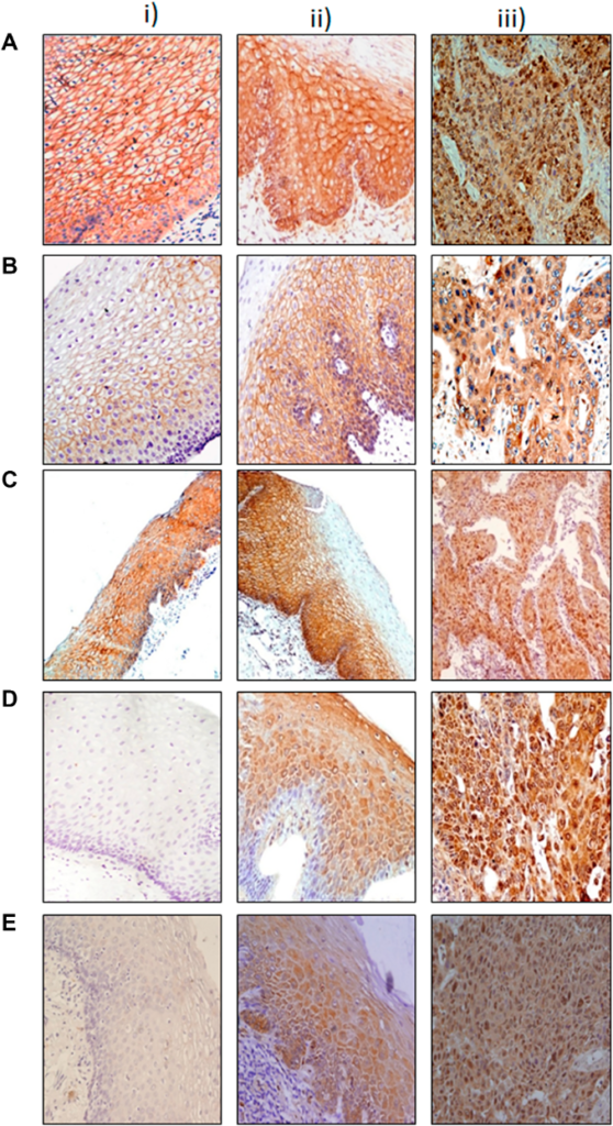
A total of 229 Ecuadorians were assessed in this study. The researchers included 155 cases of GA; 99 of the individuals with GA lived in the mountainous regions of Quito and Ambato, and 56 individuals lived in the coastal region of Guayaquil. (Controls accounted for 74 people; 25 from the coast and 49 from the mountains.) The team followed-up with all individuals in this study from 2005 to 2018 and collected blood and tissue samples. They performed immunohistochemistry and other analyses to evaluate HER2 and HIF-1 expression.

“Analyses were performed using Fisher’s exact and Breslow-Day tests for homogeneity and Kaplan-Meier curves and restricted median survival time ΔRMST.”

Results & Conclusion

After 10 years, median survival was significantly higher among GA patients living along the coast. In the GA samples, HIF-1 was observed in 66.1% of the coastal patients and in 43.4% of the mountainous patients. Positive HIF-1 expression was associated with improved survival among GA patients living in the mountains. Interestingly, in the control group (without GA), HIF-1 expression was observed in 95.9% of the mountainous samples and only 32% of the coastal samples. Their results showed that 89% of the control population exhibited gastritis.

The researchers were forthcoming about the limitations of this study. Differences in quantities and the male-to-female ratios within the GA and control coastal and mountainous groups may have influenced data. Despite these limitations, the researchers observed that coastal GA patients and individuals who expressed HIF-1 had a better prognosis. However, HIF-1 expression was only associated with better survival in the mountain region. These findings suggest that HIF-1 expression may be a protective factor against GA progression in people living at high altitudes.

“Concluding, this study suggests that HIF-1 has a differential expression pattern in gastric samples according to geographical features, being highly expressed even in non-carcinomatous cells (gastritis and normal mucosa) from individuals living in regions of high altitude, indicating that the gastric HIF-1 expression may be an adaptation of the individual to high altitudes.”

Click here to read the full research paper published by Oncotarget

ONCOTARGET VIDEOS: YouTube | LabTube | Oncotarget.com

Oncotarget is an open-access journal that publishes primarily oncology-focused research papers in a continuous publishing format. These papers are available at no cost to readers on Oncotarget.com. Open-access journals have the power to benefit humanity from the inside out by rapidly disseminating information that may be freely shared with researchers, colleagues, family, and friends around the world.

For media inquiries, please contact media@impactjournals.com.

Protein-Based Risk Model Predicts Esophageal Cancer Recurrence

Researchers developed a multi-protein expression-based risk model to predict recurrence-free survival for ESCC patients.

Listen to an audio version of this article

The Trending With Impact series highlights Oncotarget publications attracting higher visibility among readers around the world online, in the news, and on social media—beyond normal readership levels. Look for future science news about the latest trending publications here, and at Oncotarget.com.

Esophageal cancer is the sixth most common cause of death from cancer worldwide. The two main types of esophageal cancer are adenocarcinoma and esophageal squamous cell carcinoma (ESCC). ESCC arises from the cells lining the esophagus, and it is most common in areas of the world where tobacco use and alcohol consumption are high.

“Biomarkers to predict the risk of disease recurrence in Esophageal squamous cell carcinoma (ESCC) patients are urgently needed to improve treatment.”

Researchers Raghibul Hasan, Gunjan Srivastava, Akram Alyass, Rinu Sharma, Anoop Saraya, Tushar K. Chattopadhyay, Siddartha DattaGupta, Paul G. Walfish, Shyam S. Chauhan, and Ranju Ralhan from All India Institute of Medical Sciences, Mount Sinai Hospital Toronto, McMaster University, Guru Gobind Singh Indraprastha University, and the University of Toronto conducted a new study on the protein expression-based risk model they developed to predict recurrence-free survival for ESCC patients. On September 14, 2022, their research paper was published in Oncotarget’s Volume 13, and entitled, “Prediction of recurrence free survival for esophageal cancer patients using a protein signature based risk model.”

The Study

“Our study is important because: (i) it is based on changes in expression levels of the biomarker proteins in different subcellular compartments and is not limited to alterations in the overall protein expression levels; (ii) investigates the comprehensive clinical relevance of subcellular alterations in expression of multiple key components of Wnt pathway in the same ESCC patients’ cohort; (iii) correlates these findings with disease outcome and (iv) develops a Biomarker risk score for defining the risk of recurrence of ESCCs.”

Figure 1: Immunohistochemical analysis of Wnt protein in esophageal tissues.

The researchers aimed to develop and validate a panel of biomarkers with the potential to predict tumor recurrence in patients with ESCC, as well as to generate a risk model for clinical decision-making. This study enrolled 80 ESCC cases, 61 esophageal dysplastic tissues and 47 normal tissues. A multi-protein signature was generated from microarray data using the Cox proportional hazard model which was then internally validated on an independent set of samples by immunohistochemistry. The researchers demonstrated that a panel of four biomarkers (cytoplasmic β-catenin, nuclear c-Myc, nuclear DVL and membrane α-catenin) constituted the prognostic molecular signature for ESCC patients. They found that this protein signature could predict disease recurrence in patients with ESCC.

“Our panel of biomarkers predicted disease recurrence more effectively as compared to individual biomarkers analyzed in this study and demonstrated the strong predictive power of this panel of biomarkers for ESCC patients.”

Conclusion

The research team found that a panel of four biomarkers could predict disease recurrence in patients with ESCC. Furthermore, they showed that this protein signature could be used to stratify patients into high- and low-risk groups. This study provides valuable insights into the role of these proteins in the development and progression of esophageal cancer. The development of this risk model may help to tailor treatment and follow-up strategies for patients with ESCC.

“In conclusion, integrated analysis of expression of the panel of 4 proteins in ESCC patients has allowed us to validate the robustness of our biomarker panel in stratification of patients at high or low risk of disease recurrence. This risk classifier has the potential to identify the high risk patients for more rigorous personalized treatment and the low risk patients may be spared from the harmful side effects of toxic therapy as well reduce the burden on health care providers. The findings of our study set the foundations for external validation of the prognostic signature as a step forward in translation of this panel of protein markers for ESCC patients and establish their clinical relevance for larger worldwide application in future studies.”

Click here to read the full research paper published by Oncotarget

ONCOTARGET VIDEOS: YouTube | LabTube | Oncotarget.com

Oncotarget is an open-access journal that publishes primarily oncology-focused research papers in a continuous publishing format. These papers are available at no cost to readers on Oncotarget.com. Open-access journals have the power to benefit humanity from the inside out by rapidly disseminating information that may be freely shared with researchers, colleagues, family, and friends around the world.

For media inquiries, please contact media@impactjournals.com.

Immunotherapy Response Predicted by Machine Learning & Gut Microbiomes

Researchers investigated common gut microbiome features of response among immunotherapy patients with different advanced-stage cancers.

Machine Learning and Gut Microbiomes Predict Immunotherapy Response
Illustration of a gut microbiome
Listen to an audio version of this article

Immunotherapy has become a powerful breakthrough in cancer treatment, however, 50% of patients do not respond to immunotherapy. What internal or external features inhibit or confer patient or tumor response to immune system-harnessing therapeutics? These patient/tumor features that impact responsiveness to immunotherapy have yet to be fully elucidated.

“Increasing evidence has emerged that gut microbial communities help shape the host immune system [911].”

The Gut Microbiome

The gut microbiome is the collection of all microbes living in the gastrointestinal tract. A growing body of evidence indicates that the gut microbiome is an important component of the human immune system. This evidence has prompted researchers to hypothesize that the gut microbiome may play a role in immunotherapy response. It has since become a focal point in immunotherapy research and also has the potential to serve as a biomarker for immunotherapy response.

Researchers previously found evidence that specific gut bacteria can influence immunotherapy outcomes by modulating immune responses in patients with melanoma, non–small cell lung cancer and renal cell cancer. (“Treatment responders generally exhibit increased gut microbial community diversity and are enriched in certain bacterial taxa including Akkermansia and Bifidobacterium [1619].”) While this is important, it is unclear whether these response signals are generalizable across different tumor types, geographical variations in the microbiome or sequencing platforms and analysis methodologies. Identifying microbiome features associated with immunotherapy response regardless of the type of cancer or where it started in the body may be the next step toward improving immunotherapy outcomes in cancer patients.

The Study

In a new study published in Oncotarget, Hai Liang, Jay-Hyun Jo, Zhiwei Zhang, Margaret A. MacGibeny, Jungmin Han, Diana M. Proctor, Monica E. Taylor, You Che, Paul Juneau, Andrea B. Apolo, John A. McCulloch, Diwakar Davar, Hassane M. Zarour, Amiran K. Dzutsev, Isaac Brownell, Giorgio Trinchieri, James L. Gulley, and Heidi H. Kong, from the National Institutes of Health LibraryNational Cancer Institute (NCI)National Human Genome Research InstituteWest Virginia UniversityZimmerman Associates Inc., and the University of Pittsburgh, used machine learning models to analyze gut microbiomes and predict patient response to immunotherapy. On July 19, 2022, the researchers published their paper in Oncotarget’s Volume 13, entitled, “Predicting cancer immunotherapy response from gut microbiomes using machine learning models.”

To find common gut microbiome features of response to immunotherapy, a tumor-agnostic and geographically limited (U.S. patients only) approach was used in this study. The researchers’ discovery cohort included 16 patients with nine different advanced-stage cancers who were enrolled in NCI immunotherapy trials (the NCI cohort). Patient samples and 16S rRNA gene sequencing data were collected. Patients who positively responded to immunotherapies (responders) and those who did not (non-responders) were categorized. The researchers used uni- and multivariate analyses to identify common microbiome features and complex microbial community interactions. 

Common microbiome features and immunotherapy response signals in the discovery cohort were further validated with larger datasets. Three previously published 16S rRNA gene sequencing datasets from melanoma patients were added to a combined dataset. This combined dataset was used to validate the NCI cohort results in a meta-analysis. Data from all four studies were used in statistical analyses and machine learning models aimed to predict immunotherapy response.

“Using the combined dataset, we trained and validated models with machine learning algorithms to predict patients’ clinical responses, followed by cross-sequencing-platform validation using shotgun metagenomic sequencing data.”

The Results

“Results suggest baseline gut microbiome features may be predictive of clinical outcomes in oncology patients on immunotherapies, and some of these features may be generalizable across different tumor types, patient cohorts, and sequencing platforms.”

The researchers found that the gut microbiome composition of responders to immunotherapy was different from that of non-responders. Several species of bacteria were differentially abundant between responders and non-responders in the NCI cohort, and some were also consistent with results from the meta-analysis. Hierarchical clustering showed a higher immunotherapy response rate among patients with enriched bacteria in the Firmicutes phylum and a lower response rate among patients enriched in the Bacteroidetes phylum at baseline. Machine learning models using microbiome features and immunotherapy response signals found in this study demonstrated a favorable prediction accuracy with the highest AUC (area under the curve) value of around 0.75.

Conclusion

“In conclusion, analyses of our cohort and the combined microbiome dataset have provided a robust assessment of immunotherapy patients’ gut microbiomes.”

Researchers used a tumor-agnostic approach to find common gut microbiome features of response to immunotherapy in cancer patients. The machine learning models developed in this study were able to demonstrate a favorable prediction accuracy of 75% at best. These results suggest that the gut microbiome may be predictive of immunotherapy response in cancer patients and that some of these features may be generalizable across different tumor types, patient cohorts, and sequencing platforms. These findings suggest that the gut microbiome is a promising biomarker for immunotherapy response. However, more research is needed to validate these findings in larger and more diverse patient populations.

“Results suggest baseline gut microbiome features may be predictive of clinical outcomes in oncology patients on immunotherapies, and some of these features may be generalizable across different tumor types, patient cohorts, and sequencing platforms. Findings demonstrate how machine learning models can reveal microbiome-immunotherapy interactions that may ultimately improve cancer patient outcomes.”

Click here to read the full research paper published by Oncotarget

ONCOTARGET VIDEOS: YouTube | LabTube | Oncotarget.com

Oncotarget is an open-access journal that publishes primarily oncology-focused research papers in a continuous publishing format. These papers are available at no cost to readers on Oncotarget.com. Open-access journals have the power to benefit humanity from the inside out by rapidly disseminating information that may be freely shared with researchers, colleagues, family, and friends around the world.

For media inquiries, please contact media@impactjournals.com.

Epigenetics and Immunotherapy Combined Fights Rare Lymphoma

In a new Oncotarget study, researchers assessed an epigenetic and immunotherapy treatment regimen among patients with blastic mantle cell lymphoma (bMCL).

Mantle cell lymphoma
Listen to an audio version of this article

Mantle cell lymphoma (MCL) is a type of non-Hodgkin’s lymphoma (NHL) that is aggressive, difficult to treat and typically affects older adults. Recurrence and mortality rates among patients with MCL have remained high, despite recent therapeutic advances. Blastic mantle cell lymphoma (bMCL) is a rare subtype of MCL associated with a worse disease trajectory.

“Despite recent advances, MCL is incurable except with allogeneic stem cell transplant. Blastic mantle cell lymphoma (bMCL) is a rarer subtype of cMCL associated with an aggressive clinical course and poor treatment response, frequent relapse and poor outcomes.”

In previous studies, researchers reported that a combination of epigenetic and immunotherapy treatments may have synergistic activity and offer better outcomes in patients with MCL. In the current study, Francis R. LeBlanc, Zainul S. Hasanali, August Stuart, Sara Shimko, Kamal Sharma, Violetta V. Leshchenko, Samir Parekh, Haiqing Fu, Ya Zhang, Melvenia M. Martin, Mark Kester, Todd Fox, Jiangang Liao, Thomas P. Loughran, Juanita Evans, Jeffrey J. Pu, Stephen E. Spurgeon, Mirit I. Aladjem, and Elliot M. Epner from Pennsylvania State University College of MedicinePenn State Hershey Cancer InstituteWinter Haven Hospital Cassidy Cancer CenterIcahn School of Medicine at Mount SinaiNational Cancer InstituteUniversity of VirginiaUVA Cancer CenterUniversity of Arizona College of MedicineOregon Health and Science University, and Beverly Hills Cancer Center used samples from a previous trial to perform correlative studies focused on clinical results in patients with blastic MCL. On August 16, 2022, their research paper was published in Volume 13 of Oncotarget, entitled, “Combined epigenetic and immunotherapy for blastic and classical mantle cell lymphoma.”

Epigenetic and Immunotherapy

Epigenetic therapy includes a range of drugs that can target epigenetic mechanisms, including DNA methylation and posttranslational modifications of histones. For example, vorinostat (SAHA; a histone deacetylase inhibitor) and cladribine (chemotherapy that also inhibits DNA methylation) are epigenetic agents. Rituximab, a maintenance immunotherapeutic agent, is a CD20-directed monoclonal antibody. These three treatments combined encompass a novel potential epigenetic and immunotherapy treatment regimen (SCR) for mantle cell lymphoma (MCL).

“Relapsed and [treatment] naïve MCL patients were treated with vorinostat (SAHA), cladribine and rituximab (SCR) regimen and followed for OS [overall survival], progression free survival (PFS) and with correlative basic science studies to investigate potential mechanisms of action of this epigenetic/immunotherapy combination.”

The Study

Since blastic MCL patients are rare, only 13 bMCL (four relapsed, nine previously untreated) patients treated with the SCR regimen were assessed in the prospective part of this study. All patients were male and Caucasian, and the median age at diagnosis was 62 years old. The patients were treated until they achieved remission, met the criteria for removal from the study, withdrew from the study, or passed away. Four patients were changed from rituximab to ofatumumab (a potent fully-human anti-CD20 antibody) due to rituximab intolerance (allergies, reactions) or lack of efficacy.

“Of 13 bMCL patients, all patients responded to therapy, with 12 patients meeting criteria for remission (CR, n = 6; PR, n = 6). Of those achieving CR, 5 remain in CR more than 5 years after diagnosis.”

Results

After a median of 4.8 cycles of therapy, 12 patients achieved a complete response (CR), and one patient maintained stable disease (SD). The patients reported an increased overall survival greater than 40 months, and several patients maintained durable remissions without relapse for longer than five years. These results are remarkably superior to current treatment regimens with conventional chemotherapy, which range from 14.5-24 months among bMCL patients.

“The median OS of 43.4 months and PFS of 17.3 months for MCL patients with blastic disease treated with SCR therapy is one of the most important outcomes in this study.”

Another important finding was that the G/A870 CCND1 polymorphism was a strong predictor of blastic MCL, nuclear localization of cyclinD1 and response to SCR therapy. The team identified two distinct mechanisms of resistance to SCR therapy. The researchers reported that the loss of CD20 expression and evading treatment by seeking sanctuary in the central nervous system were two major resistance mechanisms to SCR therapy. 

“These data indicate that administration of epigenetic agents improves efficacy of anti-CD20 immunotherapies.”

Conclusion

Although the study sample was relatively small, the researchers’ results are promising. The SCR regimen was demonstrated to be an effective epigenetic and immunotherapy treatment for mantle cell lymphoma, with long-term remissions and improved overall survival in bMCL patients. Researchers revealed important insights into the mechanisms of action of SCR and potential resistance mechanisms. This study also highlights the potential for future research exploring the efficacy of SCR in other cancers, along with other predictive biomarkers of response.

“This approach is promising in the treatment of MCL and potentially other previously treatment refractory cancers.”

Click here to read the full research paper published by Oncotarget

ONCOTARGET VIDEOS: YouTube | LabTube | Oncotarget.com

Oncotarget is an open-access journal that publishes primarily oncology-focused research papers in a continuous publishing format. These papers are available at no cost to readers on Oncotarget.com. Open-access journals have the power to benefit humanity from the inside out by rapidly disseminating information that may be freely shared with researchers, colleagues, family, and friends around the world.

For media inquiries, please contact media@impactjournals.com.

Does a Mechanism Linking Cellular Aging to Cellular Quiescence Exist?

Researchers found that diverse geroprotectors differently affect a mechanism linking cellular aging to cellular quiescence in budding yeast.

Saccharomyces cerevisiae
Saccharomyces cerevisiae yeast budding cell under the microscope.

The Trending With Impact series highlights Oncotarget publications attracting higher visibility among readers around the world online, in the news, and on social media—beyond normal readership levels. Look for future science news about the latest trending publications here, and at Oncotarget.com.

Listen to an audio version of this article

The mechanisms of cellular aging and cellular quiescence have been preserved throughout evolution. Cellular quiescence is a temporary state of cell cycle arrest and low metabolic activity. Importantly, quiescent (Q) cells maintain the ability to quickly activate and re-enter the cell cycle (in response to the appropriate stimuli). Recent research has shown that cellular quiescence may play a role in cellular aging. 

In a 2020 study, research findings indicated that the rate at which yeast cells age is determined by a complicated program that affects 1) when a state of quiescence is entered, 2) how long quiescence is maintained and 3) when the cell exits quiescence. Researchers found that caloric restriction (CR) (a geroprotective intervention) appears to remodel this program, and this remodeling could be responsible for the CR-dependent delay of yeast chronological aging. Thus, the researchers considered the question: Does a single mechanism exist which links cellular aging to cellular quiescence? 

“We have introduced a new yeast model for studying mechanisms linking cellular aging to cellular quiescence [109110].”

In a new study, researchers (Anna Leonov, Rachel Feldman, Amanda Piano, Anthony Arlia-Ciommo, Jennifer Anne Baratang Junio, Emmanuel Orfanos, Tala Tafakori, Vicky Lutchman, Karamat Mohammad, Sarah Elsaser, Sandra Orfali, Harshvardhan Rajen, and Vladimir I. Titorenko) from Concordia University, Montreal, used a new yeast model to test their hypothesis that a mechanism exists linking cellular aging to cellular quiescence. On July 28, 2022, their research paper was published in Oncotarget and entitled, “Diverse geroprotectors differently affect a mechanism linking cellular aging to cellular quiescence in budding yeast.”

Caloric Restriction Delays Cellular Aging by Quiescence Program Changes

“Our hypothesis posits that this mechanism integrates four different processes, all of which are initiated after yeast cells cultured in a medium initially containing glucose consume it.”

In a 2017 study, researchers cultured yeast in a medium initially containing 0.2% glucose (CR). After consuming the glucose, the cells began to differentiate into quiescent and non-quiescent cell populations. Quiescent cells that developed in these cultures had different buoyant densities and could be separated into high- and low-density sub-populations. 

CR delayed yeast chronological aging by causing specific changes in four processes of a cellular quiescence program. Process one consists of a cell-cycle arrest and leads to the formation of high-density Q cells. Process two is the conversion of high-density Q cells into low-density Q cells. Processes three and four are the fast or slow decline of quiescence in low- or high-density Q cells, respectively. The researchers believe that these processes could converge into a mechanism that links cellular aging to cellular quiescence in chronologically aging budding yeast.

Figure 1: A hypothetical model for the four processes linking cellular aging to cellular quiescence. 
Figure 1: A hypothetical model for the four processes linking cellular aging to cellular quiescence. 

How do Other Geroprotectors Change the Quiescence Program?

“Here, we tested our hypothesis by assessing how four different geroprotectors influence the four processes that could link cellular aging to cellular quiescence.”

In the current study, the team’s first objective was to compare the effects of four different geroprotectors on the four quiescence processes. CR, lithocholic acid (LCA) and the single-gene deletion mutations tor1Δ and ras2Δ all delay chronological aging and extend the longevity of S. cerevisiae. Geroprotectors other than CR were examined in each of the four processes. They found that these geroprotectors differently affected processes one and two and decelerated processes three and four. Two ways of slowing down yeast chronological aging were determined by testing the four geroprotectors. One way was specific to CR and the ras2Δ mutation, and the other way was characteristic for LCA and the tor1Δ mutation.

“We selected CR and LCA to investigate the two ways different geroprotectors postpone yeast chronological aging by differently targeting the mechanism potentially linking cellular aging to cellular quiescence.”

They hypothesized that the abilities of CR and LCA to regulate the four processes of a cellular quiescence program are the same used to slow yeast chronological aging. Their next objective was to test the hypothesis that specific metabolic Q cell traits can contribute to the different effects of CR and LCA on processes one and two and their similar effects on processes three and four. Two CR-specific changes in metabolic traits of Q cells were assessed: increased intracellular concentrations of glycogen and trehalose within Q cells. 

“Therefore, we assessed the contributions of the increased intracellular concentrations of glycogen and trehalose within Q cells to the CR- and LCA-driven changes in cellular quiescence and to the CR- and LCA-promoted slowdowns of yeast chronological aging.”

Cellular Aging Delayed in Two Different Ways

In summary, study results showed that both ​​CR and the ras2Δ mutation stimulated the development of high-density Q cells (process 1) and decelerated yeast chronological aging by arresting the cell cycle in early G1, whereas LCA and the tor1Δ mutation did so by arresting the cell cycle in late G1. Both ​​CR and the ras2Δ mutation promoted an age-related conversion of high-density Q cells into low-density Q cells (process 2), whereas LCA and the tor1Δ mutation postponed this conversion. All four geroprotective interventions delayed a fast aging-associated deterioration in the quiescence of low-density Q cells (process 3) and postponed a slow aging-associated decline in the quiescence of high-density Q cells (process 4).

It is possible that the different ways these geroprotectors regulate the first two processes do not contribute to the aging-delaying capabilities of these geroprotectors. However, the researchers in this study believe there are two different ways of employing geroprotector-dependent changes in the first two processes that decelerate yeast chronological aging. They also found that a rise in trehalose within quiescent yeast contributes to chronological aging and quiescence maintenance.

“The second line of evidence for the existence of a mechanism linking cellular aging to cellular quiescence comes from our observation that an increase in intracellular trehalose within Q cells is an essential contributor to both chronological aging and quiescence maintenance in S. cerevisiae.”

Figure 6: A model for the two different ways of delaying yeast chronological aging by geroprotectors that differently affect the mechanism potentially linking cellular aging to cellular quiescence.
Figure 6: A model for the two different ways of delaying yeast chronological aging by geroprotectors that differently affect the mechanism potentially linking cellular aging to cellular quiescence.

Conclusion

“This study and our previously published data [109] provide conclusive evidence for the existence of a mechanism that links cellular aging to cellular quiescence in chronologically aging S. cerevisiae. The mechanism integrates processes 1, 2, 3 and 4 discussed above in the text and schematically depicted in Figures 1 and 6.”

Collectively, these data provide conclusive evidence for a mechanistic link between cellular aging and cellular quiescence. In the future, the researchers aim to better understand how to target the cellular quiescence program in order to delay cell aging and the onset of aging-related diseases.

“In conclusion, because the mechanisms of cellular aging and cellular quiescence are evolutionarily conserved [136266], this study makes an important next step toward the understanding of how the knowledge-based targeting of cellular quiescence can be used for slowing down cellular and organismal aging and for delaying the onset of aging-associated diseases.”

Click here to read the full research paper published by Oncotarget.

ONCOTARGET VIDEOS: YouTube | LabTube | Oncotarget.com

Oncotarget is an open-access journal that publishes primarily oncology-focused research papers in a continuous publishing format. These papers are available at no cost to readers on Oncotarget.com. Open-access journals have the power to benefit humanity from the inside out by rapidly disseminating information that may be freely shared with researchers, colleagues, family, and friends around the world.

For media inquiries, please contact media@impactjournals.com.

How Heartburn Can Turn Into Esophageal Cancer, and a Possible Biomarker

In a recent Oncotarget paper, researchers investigated telomere shortening in patients with Barrett’s esophagus as a potential biomarker of high risk for esophageal cancer.

Acid reflux / heartburn

The Trending With Impact series highlights Oncotarget publications attracting higher visibility among readers around the world online, in the news, and on social media—beyond normal readership levels. Look for future science news about the latest trending publications here, and at Oncotarget.com.

Listen to an audio version of this article

Smokers are significantly more likely than nonsmokers to have acid reflux. In many Western countries, a popular diet—known for its convenience, availability and, frankly, its lack of nutritional value—is also known to cause acid reflux. Some of the affordable foods and beverages easily accessible to Western consumers include fried food, fast foods, pizza, potato chips (and other processed snacks), high-fat meats (bacon, sausage), cheese, alcohol, soda, energy drinks, and etcetera. Unfortunately, this indulgent type of diet is accompanied by consequences beyond oily skin and an expanding waistband.

Barrett’s Esophagus

Chronic acid reflux can lead to gastroesophageal reflux disease. Gastroesophageal reflux disease can lead to Barrett’s esophagus (BE). BE is a premalignant condition in which the lining of the esophagus becomes damaged by acid reflux. BE can lead to the onset of a type of cancer called esophageal adenocarcinoma (EAC). Over the past few decades, statistics have reported that the incidence of EAC in Western populations is increasing.

“Esophageal adenocarcinoma (EAC) is on the rise in western countries with increased incidence and high mortality [12].”

Since the popularity of smoking and a heartburn-inducing diet is likely to continue in the West, the early detection of EAC is critical for improving patient outcomes. If a biomarker could indicate a BE patient’s present risk of EAC, early EAC treatment could curb incidence and mortality rates. However, such a biomarker has yet to be confirmed. On February 14, 2022, researchers from Technische Universität MünchenColumbia University Irving Medical Center and Universitätsklinikum Freiburg published the research paper, “Telomere shortening accelerates tumor initiation in the L2-IL1B mouse model of Barrett esophagus and emerges as a possible biomarker,” in Oncotarget.

“Here we aimed to provide functional evidence for the hypothesis that telomere shortening can directly contribute to tumor initiation, and thus serve as a potential biomarker for BE cancer risk stratification [2224].”

Telomere Shortening and Tumor Initiation

“Shortened telomeres is a common sight in epithelial cancers and has also been described in EAC and its precancerous lesions.”

In this study, researchers investigated the impact of shortened telomeres in a mouse model for Barrett’s esophagus (L2-IL1B). The L2-IL1B mouse model is characterized by inflammation that leads to a Barrett-like metaplasia. The team knocked out the mTERC gene (mTERC−/−), which is the catalytic subunit of telomerase in the L2-IL1B mice. 

After mTERC knockout, the researchers found that the telomeres shortened and the mice displayed signs of DNA damage. The tumor area along the squamocolumnar junction (SCJ) was increased in the second generation of these mice, and histopathological dysplasia (abnormal changes) was also increased. In vitro studies indicated that organoid formation capacity increased in BE tissue from the L2-IL1B mTERC−/− G2 mice.

“In summary, we here demonstrated a functional role of telomere shortening, a well observed property of BE, in promoting early onset esophageal tumor initiation in the L2-IL1B mouse model.”

Additional results of the study found that the telomeres in human BE epithelial cells lining the stomach with or without dysplasia were shorter than in gastric cardia tissue (the junction between the lower esophagus and the stomach). The study also found that differentiated cells that make mucus (goblet cells, which help protect the stomach lining) had longer telomeres than cells actively dividing (and more likely to become cancerous) in the columnar lined BE epithelium. 

“Moreover, besides the importance during early carcinogenesis in the mouse model, shortening of telomeres was specifically decreased in dysplastic columnar-type tissue rather than in differentiated goblet cells in human BE- and LGD tissue samples.”

Conclusion

“Here, we demonstrate that telomere dysfunction aggravates the histological phenotype, extends the tumor area in the inflammation-based L2-IL1B mouse model for BE and acts as a driver for early dysplasia development.”

In summary, these findings suggest that shortened telomeres may play a role in tumor development in a mouse model of BE and are associated with proliferating columnar epithelium in human BE. The study suggests that shortened telomeres should be evaluated further as a possible biomarker for predicting EAC cancer risk in people with BE.

“It is plausible that with our measurements we could emulate this with shortened telomeres being at higher risk of genome instability and lowered cell-to-cell variability marking clonal expansion. However, larger studies are needed to test these hypotheses.”

Click here to read the full research paper published by Oncotarget.

ONCOTARGET VIDEOS: YouTube | LabTube | Oncotarget.com

Oncotarget is an open-access journal that publishes primarily oncology-focused research papers in a continuous publishing format. These papers are available at no cost to readers on Oncotarget.com. Open-access journals have the power to benefit humanity from the inside out by rapidly disseminating information that may be freely shared with researchers, colleagues, family, and friends around the world.

For media inquiries, please contact media@impactjournals.com.

New Tool Uses NF-κB Activity to Classify HPV+ Head and Neck Cancer

Researchers developed a new tool aimed at better classifying HPV+ HNSCC patients with good or poor prognosis in an effort to personalize treatment and improve patient outcomes.

New Tool Uses NF-κB Activity to Classify HPV+ Head and Neck Cancer

Listen to an audio version of this article

Over the last 10 years in the United States, the human papillomavirus (HPV) has caused more head and neck squamous cell carcinomas (HNSCC) than uterine cervical cancers. Primarily caused either by exposure to HPV or to ethanol or tobacco, HNSCC is a disease that impairs fundamental tissues involved in respiration, speech and digestion. HPV-positive and -negative HNSCC have contrasting clinical, epidemiological and histological features. 

“A major discovery in the recent past is that HPV associated HNSCC have improved survival compared to tobacco associated tumors.”

Therefore, treating HNSCC in accordance with HPV status is crucial for avoiding unnecessarily harsh therapeutic side effects in HPV+ HNSCC patients. However, while oncologic outcomes among patients with HPV+ HNSCC are generally favorable, approximately 30% experience a more aggressive disease course and recurrence. Coupled with increasing incidence worldwide, this highlights a growing need for the development of effective clinical stratification tools to accurately identify HPV+ HNSCC patients who have a good or poor prognosis. 

In a new study, researchers—from Columbia UniversityUniversity of Illinois Cancer CenterUniversity of North Carolina at Chapel Hill, and Yale School of Medicine—developed a new tool aimed at better classifying HPV+ HNSCC patients with good or poor prognosis in an effort to personalize treatment and improve patient outcomes. Their trending research paper was published in Oncotarget on May 24, 2022, and entitled, “NF-κB over-activation portends improved outcomes in HPV-associated head and neck cancer.”

“To improve on genomic classification, we designed this study to provide a foundation for development of NF-κB related, RNA based classification strategies to better identify HPV+ HNSCC patients with good or poor prognosis that could potentially aid in future efforts towards treatment personalization.”

The Study

The researchers from this study previously found that TRAF3 and CYLD genes are negative regulators of a family of inducible transcription factors involved in inflammation, called nuclear factor kappa B or NF-κB. The team found that somatic mutations or deletions in either TRAF3 or CYLD (not commonly found in uterine cervical cancer or HPV-negative HNSCC) lead to increased NF-κB pathway activation in HPV+ HNSCC. NF-κB overactivity may lead to cancer cell growth and survival. Alterations in these NF-κB related genes may be potential therapeutic targets in HPV+ HNSCC, and their expression may be capable of predicting treatment outcomes.

“[…] we hypothesized that tumor groups based on NF-κB related gene expression may correlate with treatment outcome, considering that tumors lacking defects in TRAF3 and CYLD may have unrecognized mechanisms driving constitutive NF-κB activation.”

In the current study, the researchers developed an RNA-based NF-κB classification tool called the NF-κB Activity Classifier, or NAC. They used bioinformatics and machine learning techniques, expression-based classification, principal component (PC) analysis, gene set enrichment analysis, and weighted gene correlation network analysis (WGCNA) to verify that the NAC is indeed capable of identifying tumors with high or low NF-κB activity and tumors with good and poor survival. 

“This report validates and expands on our findings that significant expression changes related to NF-κB activity occur in the subset of HPV+ HNSCC tumors marked by TRAF3 or CYLD mutations. We are planning future studies investigating the importance of ‘long-tail’ mutations in the NF-κB pathway which might further illuminate the origins of NF-κB dysregulation in HPV+ HNSCC.”

Conclusion

“Here we present data that these subclasses may also be identified by direct assessment of NF-κB activity; as demonstrated by gene expression differences highlighted by the NF-κB Activity Classifier.”

In summary, the researchers identified genomic differences within subclasses of HPV+ HNSCC. They found that defects in TRAF3 and CYLD genes and NF-κB activity were correlated with survival. Therefore, the NF-κB Activity Classifier could be a useful guide for clinicians who make therapeutic decisions for patients with HPV+ HNSCC.

“Future applications of the NF-κB Activity Classifier may be to identify HPV+ HNSCC patients with better or worse survival with implications for treatment strategies.”

Click here to read the full research paper published by Oncotarget.

ONCOTARGET VIDEOS: YouTube | LabTube | Oncotarget.com

Oncotarget is an open-access journal that publishes primarily oncology-focused research papers in a continuous publishing format. These papers are available at no cost to readers on Oncotarget.com. Open-access journals have the power to benefit humanity from the inside out by rapidly disseminating information that may be freely shared with researchers, colleagues, family, and friends around the world.

For media inquiries, please contact media@impactjournals.com.

Trending With Impact: Dual Requirement in Stem Cell Leukemia/Lymphoma

For the first time, researchers revealed the protein interactome, phospho-proteome and total proteome for the oncogenic fusion protein BCR-FGFR1.

Figure 6: Signaling pathways activated by BCR-FGFR1.

The Trending With Impact series highlights Oncotarget publications attracting higher visibility among readers around the world online, in the news, and on social media—beyond normal readership levels. Look for future science news about the latest trending publications here, and at Oncotarget.com.

Listen to an audio version of this article

Chromosomes are found in the nucleus of cells and consist of proteins and tightly coiled strands of DNA. During cell division, chromosomal translocations can occur while the chromosomes are being copied. This type of mutation can mean that an entire chromosome has moved to another location, or that a chromosome has broken, usually into two pieces, and moved to another site. Some translocations are harmless, but others can lead to aberrant cell proliferation and cancer.

“Over the last half century, chromosomal translocations encoding functional oncogenic proteins have been identified as drivers of multiple cancers, and account for 20% of all malignant neoplasms [1, 2].”

For example, the t(8;22)(p11;q11) chromosomal translocation leads to the initiation of an oncogenic fusion protein called the Breakpoint Cluster Region Fibroblast Growth Factor Receptor 1 (BCR-FGFR1). BCR-FGFR1 is a single driver of 8p11 myeloproliferative syndrome, which is also known as stem cell leukemia/lymphoma (SCLL).

“Stem cell leukemia/lymphoma (SCLL) exhibits distinct clinical and pathological features characterized by chromosomal translocations involving the FGFR1 gene at chromosome 8p11.”

In a trending new study, researchers from the University of California San Diego and Sanford Burnham Prebys Medical Discovery Institute examined mutations in PLCγ1 and Grb2 binding sites individually and when combined together in a double mutant within BCR-FGFR1. On May 11, 2022, the research paper was published in Oncotarget and entitled, “Proteomic analysis reveals dual requirement for Grb2 and PLCγ1 interactions for BCR-FGFR1-Driven 8p11 cell proliferation.”

The Study

In this study, the researchers used quantitative proteomic analyses to identify the crucial protein-to-protein interactions that may be necessary to activate BCR-FGFR1. The team used NIH3T3, HEK293T and 32D cells to assay five types of mutations: wild type BCR-FGFR1, a kinase-dead variant of BCR-FGFR1, a derivative of BCR-FGFR1 that contained a single mutation abolishing the Grb2 interaction site, a derivative of BCR-FGFR1 that contained a single mutation abolishing the PLCγ1 interaction site, and a double mutation that abolished both interaction sites (BCR(Y177F)-FGFR1(Y766F)).

“These data demonstrate that inhibition of either signaling pathway alone fails to inhibit hematopoietic cell proliferation, and demonstrate a dual requirement for Grb2 and PLCγ1 interactions with BCR-FGFR1 for proliferation.”

When either Grb2 or PLCγ1 signaling pathway was mutated, BCR-FGFR1 activity was decreased, but never abolished. However, when both Grb2 and PLCγ1 interactions were mutated, both cell transformation and proliferation were inhibited. The team demonstrated that BCR-FGFR1 dually relies on Grb2 and PLCγ1 for biological activity and the activation of cell signaling pathways. The researchers also found that the PLCγ1 inhibitor U73122 revealed that PLCγ1 is a potential therapeutic target for BCR-FGFR1-driven hematologic malignancies. In addition, the irreversible FGFR inhibitor futibatinib suppressed downstream signaling and cell transformation. 

“We demonstrate here that BCR-FGFR1 relies dually on the small adapter protein, Grb2, and the phospholipase, PLCγ1, for biological activity and the activation of cell signaling pathways (summarized in Figure 6).”

Figure 6: Signaling pathways activated by BCR-FGFR1.

Conclusion

“Our work highlights the importance of sequencing based, mutation-specific therapies for FGFR1 induced hematologic malignancies.”

This study provides new insight into the potential molecular mechanisms underlying BCR-FGFR1 activity and identifies PLCγ1 as a therapeutic target for leukemia/lymphoma patients with this particular mutation. Future studies will be necessary to validate these findings in animal models and clinical trials. However, this study lays the groundwork for the development of new and more targeted leukemia/lymphoma therapies.

“These data unravel essential roles of Grb2 and PLCγ1 in BCR-FGFR1 mediated oncogenic growth and suggest the importance of further investigation into PLCγ1 as a potential therapeutic target in treating SCLL.”

Click here to read the full research paper published by Oncotarget.

ONCOTARGET VIDEOS: YouTube | LabTube | Oncotarget.com

Oncotarget is an open-access journal that publishes primarily oncology-focused research papers in a continuous publishing format. These papers are available at no cost to readers on Oncotarget.com. Open-access journals have the power to benefit humanity from the inside out by rapidly disseminating information that may be freely shared with researchers, colleagues, family, and friends around the world.

For media inquiries, please contact media@impactjournals.com.

Gene Variants Investigated in Polish Bladder and Kidney Cancer

Two gene variants were studied in large-scale cohorts for their potential roles in bladder and kidney cancer among Polish patients.

Genitourinary cancers are a group of cancers that affect components of the urinary tract, including the bladder and kidneys. Worldwide, bladder and kidney cancer impact men at disproportionately higher rates than women. While incidence and mortality rates of bladder cancer in most western European countries have been consistently decreasing, some countries in the region, such as Poland, have seen an increase. Bladder cancer is the 4th most common malignancy in Polish men and the 14th most common malignancy in Polish women. There is currently a need to identify more effective bladder cancer biomarkers and therapeutic targets to develop new effective treatments that improve patient outcomes.

“The association between the NOD2 c.3020insC allele and CDKN2A missense variant c.442G>A (p.P.A148T) and survival of patients with bladder or kidney cancer remains controversial.”

In April of 2022, researchers from Pomeranian Medical UniversityUniversity of Newcastle and NSW Health Pathology published the first larger-scale study in Poland to describe the clinical characteristics and survival of bladder cancer patients and kidney cancer patients associated with variants in NOD2 and CDKN2A. Their research paper was published in Oncotarget on April 22, 2022, and entitled, “Bladder cancer survival in patients with NOD2 or CDKN2A variants.”

The Study

In this study, the researchers investigated two gene variants—the NOD2 c.3020insC variant and the CDKN2A p.A148T polymorphism—and their role in bladder and kidney cancer in Polish cohorts. This NOD2 variant has been shown to occur in 7.3% of the Polish population. The CDKN2A polymorphism has been found in 3.5% of the Polish population. Therefore, these gene variants could be considered genetic risk factors for cancer. To test this hypothesis, the researchers assembled detailed participant data from a cohort of 706 bladder cancer patients and 410 kidney cancer patients. The team compiled control data from over 5,000 unselected, cancer-free individuals.

“To our knowledge, this is the first larger-scale study describing the clinical characteristics and survival of bladder and kidney cancer patients that is associated with the NOD2 c.3020insC allele and the CDKN2A p.A148T polymorphism in Poland.”

After performing the variant analysis in the cohort of Polish patients with bladder cancer, the team found that 8.9% of these patients carried the NOD2 variant and 5.2% carried the CDKN2A variant. However, their analysis revealed that neither the NOD2 nor the CDKN2A variant played a significant role in the survival of patients with bladder cancer. In performing the variant analysis in the cohort of Polish patients with kidney cancer, they found that 7.3% of these patients carried the NOD2 variant and 3.4% carried the CDKN2A variant. The researchers did not observe any statistically significant relationship between kidney cancer and either variant. However, they were not able to perform a survival analysis in the kidney cancer cohort.

Conclusion

The researchers found that the NOD2 c.3020insC variant and the CDKN2A p.A148T polymorphism were not significantly associated with the survival of bladder cancer patients, regardless of age, cancer family history, smoking status, and sex. To date, this is the first larger-scale study to examine these variants in association with clinical characteristics and survival of Polish patients with bladder cancer.

“In summary, the results of this study indicate that neither the NOD2 c.3020insC variant or the CDKN2A p.A148T polymorphism are associated with the survival of bladder cancer patients regardless of age, cancer family history, smoking status, and sex. Thus, the NOD2 c.3020insC or the CDKN2A p.A148T polymorphism cannot be added to the list of genes that are associated with an increased susceptibility to bladder or kidney cancer at this time.”

Click here to read the full research paper published by Oncotarget.

ONCOTARGET VIDEOS: YouTube | LabTube | Oncotarget.com

Oncotarget is an open-access journal that publishes primarily oncology-focused research papers in a continuous publishing format. These papers are available at no cost to readers on Oncotarget.com. Open-access journals have the power to benefit humanity from the inside out by rapidly disseminating information that may be freely shared with researchers, colleagues, family, and friends around the world.

For media inquiries, please contact media@impactjournals.com.

Trending With Impact: Analysis of Mutational Burden in NSCLC

Researchers conducted a multi-site cohort study of tumor mutational burden among hundreds of patients diagnosed with stage IV non-small cell lung cancer (NSCLC).

Lung cancer x-ray
Lung cancer x-ray

The Trending With Impact series highlights Oncotarget publications attracting higher visibility among readers around the world online, in the news, and on social media—beyond normal readership levels. Look for future science news about the latest trending publications here, and at Oncotarget.com.

Listen to an audio version of this article

While a high tumor mutational burden (TMB) may seem unfavorable in the midst of battling non-small cell lung cancer (NSCLC), a higher TMB has been associated with a higher number of neoantigens. The presence of more neoantigens can potentially elicit a stronger immune response. Therefore, TMB may be a viable biomarker of tumor response to immunotherapeutic agents. However, the definitions, parameters and units used to measure high- and low-TMB have been inconsistent over the years. Today, the consensus unit for reporting TMB has shifted to mutations per megabase (mut/Mb). The common cut-off for high- vs. low-TMB from tissue samples is >10 mut/Mb in NSCLC.

“Despite inconsistencies with TMB definition and reporting over time, high TMB has consistently been associated with improved clinical benefit among patients receiving immunotherapy for NSCLC [22].”

Researchers—from University of UtahUniversity of Minnesota DuluthHuntsman Cancer InstituteH. Lee Moffitt Cancer Center and Research InstituteBaptist Health Medical GroupMetroHealth Medical CenterRutgers Cancer Institute of New JerseyUniversity of Southern CaliforniaSaint Luke’s Cancer InstituteUniversity of Kentucky, and Bristol Myers Squibb—used the newest consensus unit and common cut-off parameters for TMB expression to measure TMB’s relationship to treatment response and survival outcomes among metastatic NSCLC patients. Their trending research paper was published in Oncotarget’s Volume 13 on January 31, 2022, and entitled, “Real-world survival analysis by tumor mutational burden in non-small cell lung cancer: a multisite U.S. study.”

“The purpose of this study is to evaluate clinical outcomes by TMB among NSCLC patients treated with immunotherapy containing regimens in the first-line setting.”

The Study

Participants in this large cohort study included 667 patients who had been diagnosed with stage IV NSCLC and treated with any NSCLC-related treatment. Patients were recruited from nine different academic and community cancer centers across the United States. The researchers intended to utilize this “real-world” dataset and hoped it would allow them to realistically assess the role of TMB as a potential biomarker of NSCLC response to treatment.

First, the team collected demographic and clinical characteristics and separated them into two groups: TMB greater or less than 10 mut/Mb. Characteristics included age, sex, race, body mass index, smoking history, PD-L1 expression, comorbidities, Eastern Cooperative Oncology Group performance status (ECOG PS) at diagnosis, histology subtype, Stage at metastatic diagnosis, and site of metasteses. Interestingly, a history of smoking was significantly associated with a TMB greater than 10 mut/Mb.

“Smoking status was significantly associated with TMB >10 with 91% of patients reported as current or former smokers compared to 61% in the TMB <10 cohort (p < 0.01, Table 1).”

The Results

The researchers found no association between TMB and age, PD-L1 expression, tumor histology, or cancer stage at diagnosis. Next, the team assessed for significant associations between TMB and 17 genomic alterations. They found that lower TMB was associated with ALK and EGFR alterations. Higher TMB was associated with TP53 alterations. The researchers investigated the association between TMB and treatment patterns and responses. The overall response rate was very similar in both groups. 

A multivariable model was used to analyze overall patient survival and progression-free survival (PFS) for first-line immunotherapy containing regimens based on TMB. The model controlled for the initial patient characteristics and did not demonstrate significantly different results for overall survival in the two groups. However, the researchers found in a subgroup analysis that, of the patients who received TMB testing within 60 days of receiving immunotherapy treatment, those with TMB >10 demonstrated significantly longer overall survival compared to their TMB <10 counterparts. In terms of PFS, they found that PFS was longer among patients with TMB >10 in the cohort and subgroup analyses. PFS was significantly longer when treated with an immunotherapy-containing regimen first-line compared to a first-line treatment of chemotherapy. An association between TMB and PD-L1 expression was not found in this study.

Conclusion

“This study evaluated two broad questions: (1) The distribution of TMB in the real world and its association with baseline clinical and demographic features (n = 677) and (2) the association between TMB and clinical outcomes among NSCLC patients who received first-line immunotherapy (n = 224).”

Results of the study confirmed the association between a higher TMB and smoking history, as well as the benefits of first-line immunotherapy within two months of TMB testing. While the researchers were forthcoming about limitations in their study, metastatic NSCLC patients with TMB>10 who were treated with first-line immunotherapy had improved overall survival and progression-free survival.

“Based on the results in this study and prior research, TMB along with other biomarkers, such as PD-L1, may help identify patients more likely to benefit from first-line immunotherapy.”

Click here to read the full research paper published by Oncotarget.

ONCOTARGET VIDEOS: YouTube | LabTube | Oncotarget.com

Oncotarget is an open-access journal that publishes primarily oncology-focused research papers in a continuous publishing format. These papers are available at no cost to readers on Oncotarget.com. Open-access journals have the power to benefit humanity from the inside out by rapidly disseminating information that may be freely shared with researchers, colleagues, family, and friends around the world.

For media inquiries, please contact media@impactjournals.com.