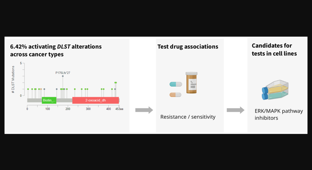
The increased expression of DLST has recently been associated with increased tumor aggression and a poor prognosis in neuroblastoma and triple-negative breast cancer.
Figure 3: Analysis of DLST-activated cell lines revealed sensitivity to protein kinase inhibiting the ERK/MAPK pathway.
The Trending With Impact series highlights Oncotarget publications attracting higher visibility among readers around the world online, in the news, and on social media—beyond normal readership levels. Look for future science news about the latest trending publications here, and at Oncotarget.com.
—
Listen to an audio version of this article
Dihydrolipoamide S-succinyltransferase (DLST) is a crucial gene/protein/enzyme involved in the oxidative phosphorylation (OXPHOS) pathway and cellular energy production. Recent studies have demonstrated that, in neuroblastoma and triple-negative breast cancer (TNBC), increased expression of DLST is associated with increased tumor aggression and a poor disease prognosis. Researchers also found that, in leukemia and TNBC cell lines, the knockdown of DLST leads to apoptosis. These findings suggest that neuroblastoma and TNBC may benefit from DLST-inhibiting cancer therapy.
“With the advent of complex genetic datasets of roughly 1000 cell lines in the Cancer Cell Line Encyclopedia (CCLE) and on drug resistance in the Genomics of Drug Sensitivity in Cancer project (GDSC), analyses of drug sensitivity have become possible on a larger scale [6, 7].”
Since neuroblastoma and TNBC tumor cell viability may be DLST-dependent, DLST is a promising target for cancer therapy. The researchers used the Cancer Cell Line Encyclopedia (CCLE) to identify cancer cell lines with DLST amplifications or high mRNA levels. They then measured the sensitivity of these DLST+ cell lines to 250 drugs in the GDSC dataset and compared the data to a subset of cell lines without DLST amplifications or high mRNA levels.
“To identify drugs that inhibit viability of specifically DLST-activated tumor cells, we compared cell lines with supposedly activating changes of DLST (DNA amplification, high mRNA levels) to cell lines without DLST changes.”
Results & Conclusions
“DLST-altered cell lines were more sensitive to 7 approved drugs, among these obatoclax mesylate, a BCL2 inhibitor that reduces OXPHOS in human leukemia stem cells.”
The researchers identified seven drug candidates that demonstrated significantly higher sensitivity in DLST+ cell lines than in the control cell lines. In addition to a BCL2 inhibitor found to reduce OXPHOS, multiple protein kinase inhibitors were identified as efficient in the DLST+ cell lines. This suggests that DLST-altered cell lines may also be vulnerable to ERK/MAPK pathway-targeting drugs. The researchers propose that the drug candidates identified in this study warrant further drug efficacy testing in knock-in cell lines and DLST-activated tumors.
“We therefore conclude that, in addition to OXPHOS, protein kinases could be potential targets of therapy in the presence of DLST amplifications or high mRNA levels.”
Click hereto read the full research paper published by Oncotarget.
Oncotarget is an open-access, peer reviewed journal that has published primarily oncology-focused research papers since 2010. These papers are available to readers (at no cost and free of subscription barriers) in a continuous publishing format at Oncotarget.com. Oncotarget is indexed/archived on MEDLINE / PMC / PubMed.
In a new study, researchers investigated the plasma growth hormone as a potential biomarker of response to atezolizumab and bevacizumab in advanced hepatocellular carcinoma patients.
The Trending With Impact series highlights Oncotarget publications attracting higher visibility among readers around the world online, in the news, and on social media—beyond normal readership levels. Look for future science news about the latest trending publications here, and at Oncotarget.com.
—
Listen to an audio version of this article
Hepatocellular carcinoma (HCC) is a highly aggressive cancer of the liver with a very poor prognosis; many patients pass away within a year of diagnosis. Currently, there is no effective screening method for HCC and thus, 80% of patients are diagnosed at advanced stages. This makes treatment difficult and often unsuccessful. As a result, new treatments for HCC are constantly being explored.
“This study investigated the biomarker value of plasma growth hormone (GH) level as a potential biomarker to predict outcome in unresectable HCC patients treated with current standard therapy, atezolizumab plus bevacizumab (Atezo/Bev).”
The Study
Plasma growth hormone (GH) is a potential biomarker that had not previously been evaluated in relation to this treatment regimen in HCC before. In this study, the researchers included 37 patients with advanced HCC. The patients received atezolizumab plus bevacizumab at the MD Anderson Cancer Center between June 2018 and November 2021. The median age of the patients was 67 years old, and the vast majority were male (83.8%).
The team measured plasma GH levels, progression-free survival (PFS) and overall survival (OS). Plasma GH levels were measured by ELISA and used to stratify the HCC patients into GH-high and GH-low groups. The Kaplan-Meier method was used to calculate median OS and PFS. The Log-rank test was used to compare survival outcomes between GH-high and -low groups.
“To the best of our knowledge, this is the first study to assess clinical prognostic value of plasma GH level in patients who have received atezolizumab plus bevacizumab in clinical setting.”
The results of the study showed that plasma GH levels significantly correlated with OS. At the time of the analysis, the one-year survival rate was 70% among GH-low patients and 33% among GH-high patients. OS was significantly superior in GH-low compared to GH-high patients. PFS showed a non-significant trend in favor of GH-low patients compared to the GH-high group.
Conclusion
“Despite the small data size, plasma GH levels were strongly predictive of the disease outcome in patients treated with Atezo/Bev.”
The study concluded that plasma GH levels may be a promising biomarker for predicting response to atezolizumab plus bevacizumab in advanced HCC patients. Further, plasma GH levels may be used to stratify advanced HCC patients into high- and low-risk groups. The researchers recommend further research in larger scale and different populations to validate the findings and explore plasma GH levels as a potential biomarker of response to this treatment regimen in HCC.
“In conclusion, our study demonstrate[s] that plasma GH represents a candidate biomarker for predicting treatment outcomes in patients with advanced HCC treated with Atezo/Bev. Future studies in larger randomized clinical trial and with a more diverse ethnic, race, and gender background are warranted to further validate these findings.”
Click here to read the full research paper published by Oncotarget.
Oncotarget is an open-access journal that publishes primarily oncology-focused research papers in a continuous publishing format. These papers are available at no cost to readers on Oncotarget.com. Open-access journals have the power to benefit humanity from the inside out by rapidly disseminating information that may be freely shared with researchers, colleagues, family, and friends around the world.
In this trending new study, researchers used CRISPR-based genome-wide screens to identify genetic determinants of PARP10-mediated cellular survival.
Listen to an audio version of this article
The Trending With Impact series highlights Oncotarget publications attracting higher visibility among readers around the world online, in the news, and on social media—beyond normal readership levels. Look for future science news about the latest trending publications here, and at Oncotarget.com.
—
Genetic interactions involved in the survival of cancer cells are potential therapeutic targets in personalized cancer therapy. “Synthetic lethal” is a type of genetic interaction where the knockout of one gene can cause cell death but only in the presence of another dependent gene. Cancer researchers view synthetic lethality screening as a powerful tool in precision medicine.
“Identifying genetic susceptibilities based on PARP10 expression levels is thus potentially relevant for finding new targets for precision oncology.”
“Here, we employed complementary CRISPR loss-of-function genome-wide screening to identify genes required for proliferation of PARP10-overexpressing and PARP10-knockout cells.”
The Study
To identify potential synthetic lethal targets, the researchers conducted a CRISPR-based, genome-wide genetic screen of both PARP10-overexpressing and PARP10-knockout tumorigenic and non-tumorigenic breast cells. The screen looked for genes that were required for cell proliferation in the presence of PARP10 overexpression or PARP10 knockout.
“Here, we performed a series of CRISPR genome-wide loss-of-function screens in isogenic control and PARP10-overexpressing or PARP10-knockout cell lines, to identify genetic determinants of PARP10-mediated cellular survival.”
In the PARP10 overexpressing cells, the top results from their CRISPR screen were validated with biological pathway enrichment analyses, using both KEGG and Gene Ontology databases. A functional interaction between ATM and PARP10 expression was found. ATM promoted cell proliferation in PARP10-overexpressing cells.
In the genome-wide CRISPR knockout screens, genes required for the viability of PARP10-knockout cells were identified. In the PARP10 knockout cells, the top results from their CRISPR screen were validated with biological pathway enrichment analyses, using both KEGG and Gene Ontology databases. They identified the CDK2-Cyclin E1 complex as a genetic determinant for the proliferation of PARP10-knockout cells.
“Our work identifies a network of functionally relevant PARP10 synthetic interactions, and reveals a set of factors which can potentially be targeted in personalized cancer therapy.”
Conclusion
The researchers identified several genes that were differentially required for cell proliferation in the presence of PARP10 overexpression or knockout. Some of these genes have been previously implicated in cancer, while others were novel candidate cancer targets. The identification of these potential synthetic lethal interactions provides new insights into the role of PARP10 in cancer and may be useful for precision oncology. This study highlights the importance of using complementary CRISPR-based screens to identify potential cancer targets.
“We found that DNA repair factors, including ATM, a master regulator of the DNA damage checkpoint response, are specifically promoting the proliferation of PARP10-overexpressing cells. Moreover, we identified a role for PARP10 in regulating ATM recruitment to stressed replication forks. Finally, we found that the CDK2-cyclin E1 complex is specifically required for the proliferation of PARP10-deficient cells. Our work reveals novel PARP10 genetic interactions of functional relevance and identifies a set of factors which can potentially be targeted in personalized cancer therapy.”
Click here to read the full research paper published by Oncotarget.
Oncotarget is an open-access journal that publishes primarily oncology-focused research papers in a continuous publishing format. These papers are available at no cost to readers on Oncotarget.com. Open-access journals have the power to benefit humanity from the inside out by rapidly disseminating information that may be freely shared with researchers, colleagues, family, and friends around the world.
Researchers developed a multi-protein expression-based risk model to predict recurrence-free survival for ESCC patients.
Listen to an audio version of this article
The Trending With Impact series highlights Oncotarget publications attracting higher visibility among readers around the world online, in the news, and on social media—beyond normal readership levels. Look for future science news about the latest trending publications here, and at Oncotarget.com.
—
Esophageal cancer is the sixth most common cause of death from cancer worldwide. The two main types of esophageal cancer are adenocarcinoma and esophageal squamous cell carcinoma (ESCC). ESCC arises from the cells lining the esophagus, and it is most common in areas of the world where tobacco use and alcohol consumption are high.
“Biomarkers to predict the risk of disease recurrence in Esophageal squamous cell carcinoma (ESCC) patients are urgently needed to improve treatment.”
“Our study is important because: (i) it is based on changes in expression levels of the biomarker proteins in different subcellular compartments and is not limited to alterations in the overall protein expression levels; (ii) investigates the comprehensive clinical relevance of subcellular alterations in expression of multiple key components of Wnt pathway in the same ESCC patients’ cohort; (iii) correlates these findings with disease outcome and (iv) develops a Biomarker risk score for defining the risk of recurrence of ESCCs.”
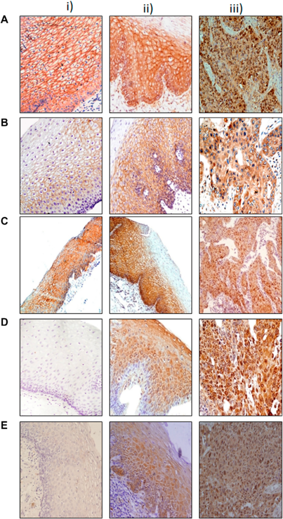
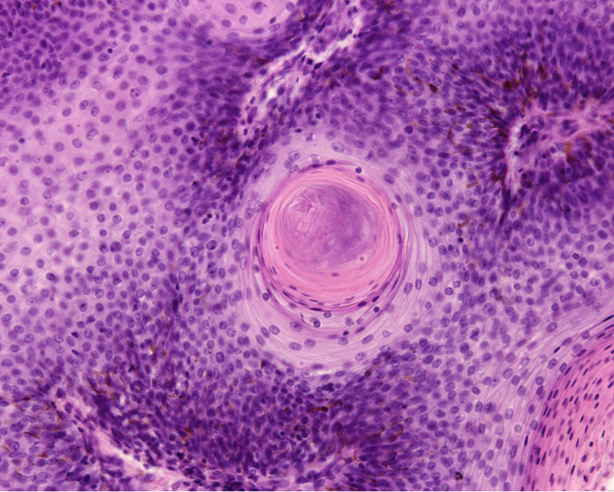
Figure 1: Immunohistochemical analysis of Wnt protein in esophageal tissues.
The researchers aimed to develop and validate a panel of biomarkers with the potential to predict tumor recurrence in patients with ESCC, as well as to generate a risk model for clinical decision-making. This study enrolled 80 ESCC cases, 61 esophageal dysplastic tissues and 47 normal tissues. A multi-protein signature was generated from microarray data using the Cox proportional hazard model which was then internally validated on an independent set of samples by immunohistochemistry. The researchers demonstrated that a panel of four biomarkers (cytoplasmic β-catenin, nuclear c-Myc, nuclear DVL and membrane α-catenin) constituted the prognostic molecular signature for ESCC patients. They found that this protein signature could predict disease recurrence in patients with ESCC.
“Our panel of biomarkers predicted disease recurrence more effectively as compared to individual biomarkers analyzed in this study and demonstrated the strong predictive power of this panel of biomarkers for ESCC patients.”
Conclusion
The research team found that a panel of four biomarkers could predict disease recurrence in patients with ESCC. Furthermore, they showed that this protein signature could be used to stratify patients into high- and low-risk groups. This study provides valuable insights into the role of these proteins in the development and progression of esophageal cancer. The development of this risk model may help to tailor treatment and follow-up strategies for patients with ESCC.
“In conclusion, integrated analysis of expression of the panel of 4 proteins in ESCC patients has allowed us to validate the robustness of our biomarker panel in stratification of patients at high or low risk of disease recurrence. This risk classifier has the potential to identify the high risk patients for more rigorous personalized treatment and the low risk patients may be spared from the harmful side effects of toxic therapy as well reduce the burden on health care providers. The findings of our study set the foundations for external validation of the prognostic signature as a step forward in translation of this panel of protein markers for ESCC patients and establish their clinical relevance for larger worldwide application in future studies.”
Click here to read the full research paper published by Oncotarget.
Oncotarget is an open-access journal that publishes primarily oncology-focused research papers in a continuous publishing format. These papers are available at no cost to readers on Oncotarget.com. Open-access journals have the power to benefit humanity from the inside out by rapidly disseminating information that may be freely shared with researchers, colleagues, family, and friends around the world.
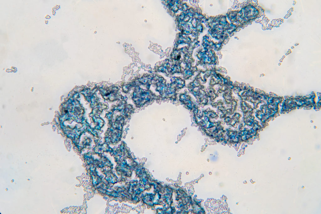
Researchers found that diverse geroprotectors differently affect a mechanism linking cellular aging to cellular quiescence in budding yeast.
Saccharomyces cerevisiae yeast budding cell under the microscope.
The Trending With Impact series highlights Oncotarget publications attracting higher visibility among readers around the world online, in the news, and on social media—beyond normal readership levels. Look for future science news about the latest trending publications here, and at Oncotarget.com.
—
Listen to an audio version of this article
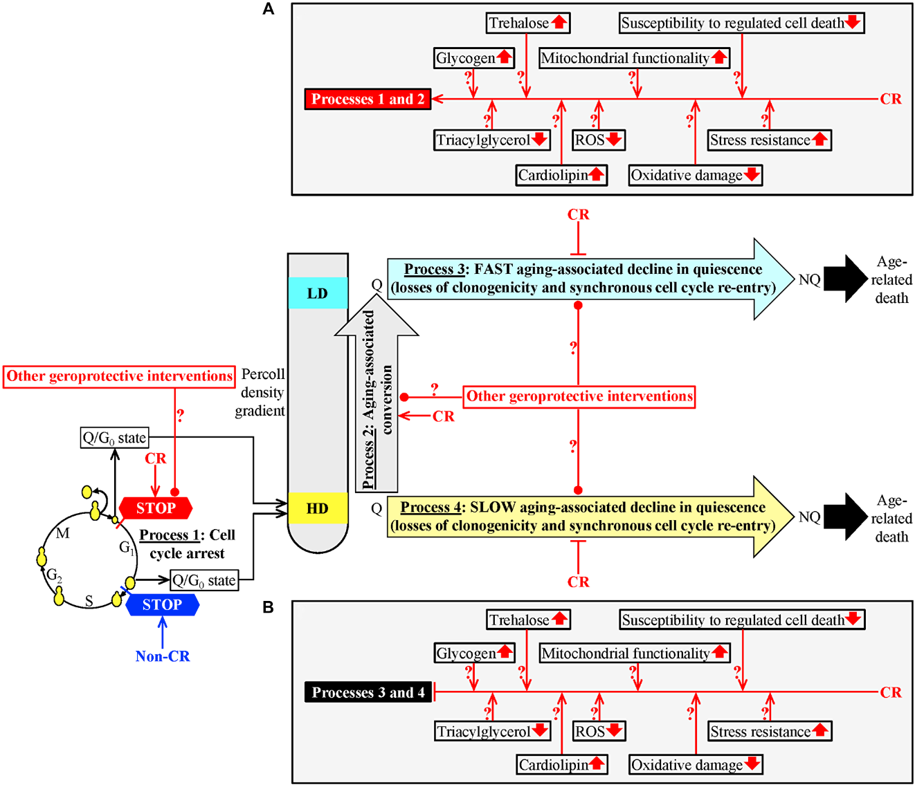
The mechanisms of cellular aging and cellular quiescence have been preserved throughout evolution. Cellular quiescence is a temporary state of cell cycle arrest and low metabolic activity. Importantly, quiescent (Q) cells maintain the ability to quickly activate and re-enter the cell cycle (in response to the appropriate stimuli). Recent research has shown that cellular quiescence may play a role in cellular aging.
In a 2020 study, research findings indicated that the rate at which yeast cells age is determined by a complicated program that affects 1) when a state of quiescence is entered, 2) how long quiescence is maintained and 3) when the cell exits quiescence. Researchers found that caloric restriction (CR) (a geroprotective intervention) appears to remodel this program, and this remodeling could be responsible for the CR-dependent delay of yeast chronological aging. Thus, the researchers considered the question: Does a single mechanism exist which links cellular aging to cellular quiescence?
“We have introduced a new yeast model for studying mechanisms linking cellular aging to cellular quiescence [109, 110].”
In a new study, researchers (Anna Leonov, Rachel Feldman, Amanda Piano, Anthony Arlia-Ciommo, Jennifer Anne Baratang Junio, Emmanuel Orfanos, Tala Tafakori, Vicky Lutchman, Karamat Mohammad, Sarah Elsaser, Sandra Orfali, Harshvardhan Rajen, and Vladimir I. Titorenko) from Concordia University, Montreal, used a new yeast model to test their hypothesis that a mechanism exists linking cellular aging to cellular quiescence. On July 28, 2022, their research paper was published in Oncotarget and entitled, “Diverse geroprotectors differently affect a mechanism linking cellular aging to cellular quiescence in budding yeast.”
Caloric Restriction Delays Cellular Aging by Quiescence Program Changes
“Our hypothesis posits that this mechanism integrates four different processes, all of which are initiated after yeast cells cultured in a medium initially containing glucose consume it.”
In a 2017 study, researchers cultured yeast in a medium initially containing 0.2% glucose (CR). After consuming the glucose, the cells began to differentiate into quiescent and non-quiescent cell populations. Quiescent cells that developed in these cultures had different buoyant densities and could be separated into high- and low-density sub-populations.
CR delayed yeast chronological aging by causing specific changes in four processes of a cellular quiescence program. Process one consists of a cell-cycle arrest and leads to the formation of high-density Q cells. Process two is the conversion of high-density Q cells into low-density Q cells. Processes three and four are the fast or slow decline of quiescence in low- or high-density Q cells, respectively. The researchers believe that these processes could converge into a mechanism that links cellular aging to cellular quiescence in chronologically aging budding yeast.
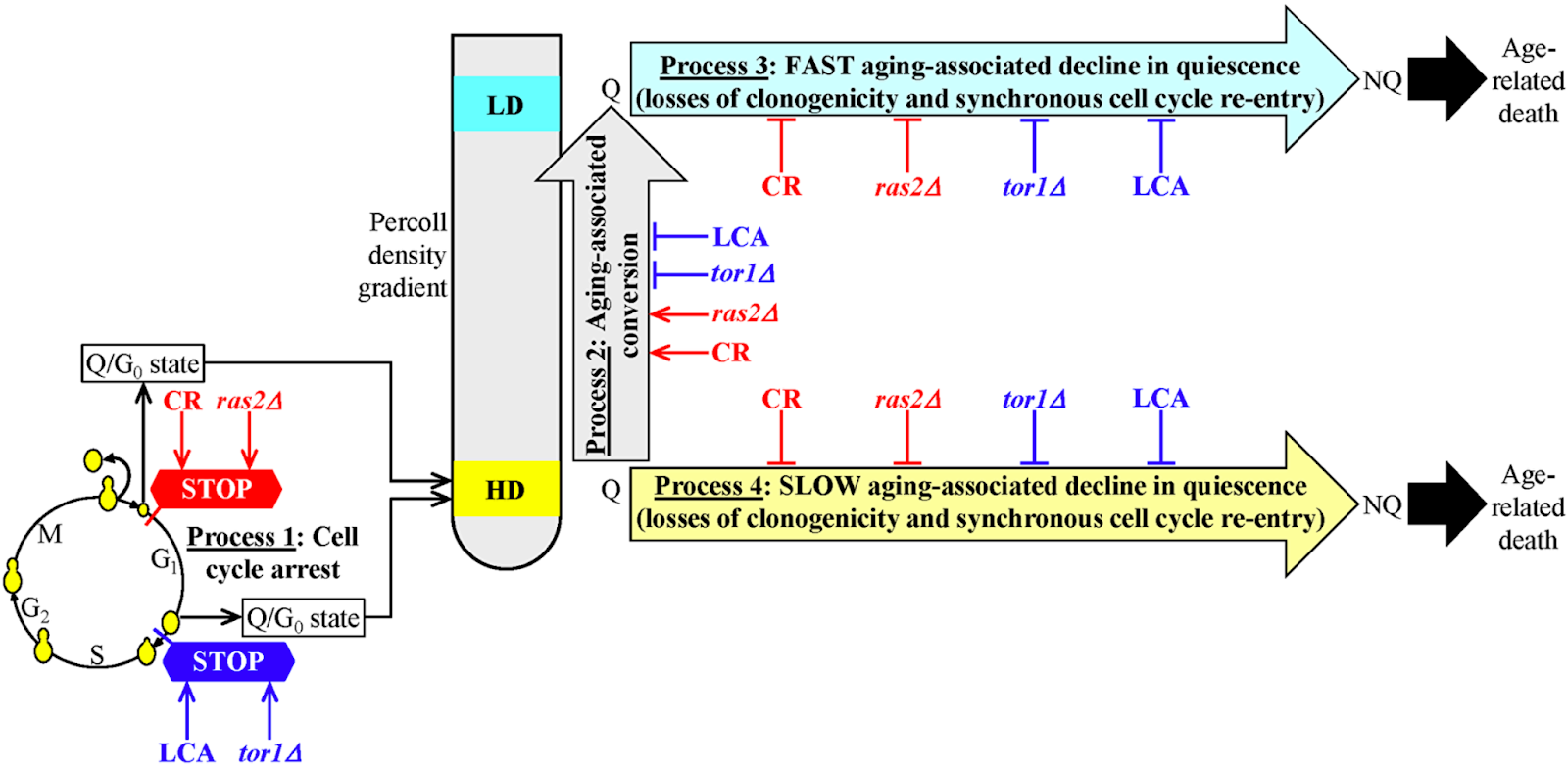
Figure 1: A hypothetical model for the four processes linking cellular aging to cellular quiescence.
How do Other Geroprotectors Change the Quiescence Program?
“Here, we tested our hypothesis by assessing how four different geroprotectors influence the four processes that could link cellular aging to cellular quiescence.”
In the current study, the team’s first objective was to compare the effects of four different geroprotectors on the four quiescence processes. CR, lithocholic acid (LCA) and the single-gene deletion mutations tor1Δ and ras2Δ all delay chronological aging and extend the longevity of S. cerevisiae. Geroprotectors other than CR were examined in each of the four processes. They found that these geroprotectors differently affected processes one and two and decelerated processes three and four. Two ways of slowing down yeast chronological aging were determined by testing the four geroprotectors. One way was specific to CR and the ras2Δ mutation, and the other way was characteristic for LCA and the tor1Δ mutation.
“We selected CR and LCA to investigate the two ways different geroprotectors postpone yeast chronological aging by differently targeting the mechanism potentially linking cellular aging to cellular quiescence.”
They hypothesized that the abilities of CR and LCA to regulate the four processes of a cellular quiescence program are the same used to slow yeast chronological aging. Their next objective was to test the hypothesis that specific metabolic Q cell traits can contribute to the different effects of CR and LCA on processes one and two and their similar effects on processes three and four. Two CR-specific changes in metabolic traits of Q cells were assessed: increased intracellular concentrations of glycogen and trehalose within Q cells.
“Therefore, we assessed the contributions of the increased intracellular concentrations of glycogen and trehalose within Q cells to the CR- and LCA-driven changes in cellular quiescence and to the CR- and LCA-promoted slowdowns of yeast chronological aging.”
Cellular Aging Delayed in Two Different Ways
In summary, study results showed that both CR and the ras2Δ mutation stimulated the development of high-density Q cells (process 1) and decelerated yeast chronological aging by arresting the cell cycle in early G1, whereas LCA and the tor1Δ mutation did so by arresting the cell cycle in late G1. Both CR and the ras2Δ mutation promoted an age-related conversion of high-density Q cells into low-density Q cells (process 2), whereas LCA and the tor1Δ mutation postponed this conversion. All four geroprotective interventions delayed a fast aging-associated deterioration in the quiescence of low-density Q cells (process 3) and postponed a slow aging-associated decline in the quiescence of high-density Q cells (process 4).
It is possible that the different ways these geroprotectors regulate the first two processes do not contribute to the aging-delaying capabilities of these geroprotectors. However, the researchers in this study believe there are two different ways of employing geroprotector-dependent changes in the first two processes that decelerate yeast chronological aging. They also found that a rise in trehalose within quiescent yeast contributes to chronological aging and quiescence maintenance.
“The second line of evidence for the existence of a mechanism linking cellular aging to cellular quiescence comes from our observation that an increase in intracellular trehalose within Q cells is an essential contributor to both chronological aging and quiescence maintenance in S. cerevisiae.”
Figure 6: A model for the two different ways of delaying yeast chronological aging by geroprotectors that differently affect the mechanism potentially linking cellular aging to cellular quiescence.
Conclusion
“This study and our previously published data [109] provide conclusive evidence for the existence of a mechanism that links cellular aging to cellular quiescence in chronologically aging S. cerevisiae. The mechanism integrates processes 1, 2, 3 and 4 discussed above in the text and schematically depicted in Figures 1 and 6.”
Collectively, these data provide conclusive evidence for a mechanistic link between cellular aging and cellular quiescence. In the future, the researchers aim to better understand how to target the cellular quiescence program in order to delay cell aging and the onset of aging-related diseases.
“In conclusion, because the mechanisms of cellular aging and cellular quiescence are evolutionarily conserved [1, 3, 62, 66], this study makes an important next step toward the understanding of how the knowledge-based targeting of cellular quiescence can be used for slowing down cellular and organismal aging and for delaying the onset of aging-associated diseases.”
Click hereto read the full research paper published by Oncotarget.
Oncotarget is an open-access journal that publishes primarily oncology-focused research papers in a continuous publishing format. These papers are available at no cost to readers on Oncotarget.com. Open-access journals have the power to benefit humanity from the inside out by rapidly disseminating information that may be freely shared with researchers, colleagues, family, and friends around the world.
In a recent Oncotarget paper, researchers investigated telomere shortening in patients with Barrett’s esophagus as a potential biomarker of high risk for esophageal cancer.
The Trending With Impact series highlights Oncotarget publications attracting higher visibility among readers around the world online, in the news, and on social media—beyond normal readership levels. Look for future science news about the latest trending publications here, and at Oncotarget.com.
—
Listen to an audio version of this article
Smokers are significantly more likely than nonsmokers to have acid reflux. In many Western countries, a popular diet—known for its convenience, availability and, frankly, its lack of nutritional value—is also known to cause acid reflux. Some of the affordable foods and beverages easily accessible to Western consumers include fried food, fast foods, pizza, potato chips (and other processed snacks), high-fat meats (bacon, sausage), cheese, alcohol, soda, energy drinks, and etcetera. Unfortunately, this indulgent type of diet is accompanied by consequences beyond oily skin and an expanding waistband.
Barrett’s Esophagus
Chronic acid reflux can lead to gastroesophageal reflux disease. Gastroesophageal reflux disease can lead to Barrett’s esophagus (BE). BE is a premalignant condition in which the lining of the esophagus becomes damaged by acid reflux. BE can lead to the onset of a type of cancer called esophageal adenocarcinoma (EAC). Over the past few decades, statistics have reported that the incidence of EAC in Western populations is increasing.
“Esophageal adenocarcinoma (EAC) is on the rise in western countries with increased incidence and high mortality [1, 2].”
“Here we aimed to provide functional evidence for the hypothesis that telomere shortening can directly contribute to tumor initiation, and thus serve as a potential biomarker for BE cancer risk stratification [22, 24].”
Telomere Shortening and Tumor Initiation
“Shortened telomeres is a common sight in epithelial cancers and has also been described in EAC and its precancerous lesions.”
In this study, researchers investigated the impact of shortened telomeres in a mouse model for Barrett’s esophagus (L2-IL1B). The L2-IL1B mouse model is characterized by inflammation that leads to a Barrett-like metaplasia. The team knocked out the mTERC gene (mTERC−/−), which is the catalytic subunit of telomerase in the L2-IL1B mice.
After mTERC knockout, the researchers found that the telomeres shortened and the mice displayed signs of DNA damage. The tumor area along the squamocolumnar junction (SCJ) was increased in the second generation of these mice, and histopathological dysplasia (abnormal changes) was also increased. In vitro studies indicated that organoid formation capacity increased in BE tissue from the L2-IL1B mTERC−/− G2 mice.
“In summary, we here demonstrated a functional role of telomere shortening, a well observed property of BE, in promoting early onset esophageal tumor initiation in the L2-IL1B mouse model.”
Additional results of the study found that the telomeres in human BE epithelial cells lining the stomach with or without dysplasia were shorter than in gastric cardia tissue (the junction between the lower esophagus and the stomach). The study also found that differentiated cells that make mucus (goblet cells, which help protect the stomach lining) had longer telomeres than cells actively dividing (and more likely to become cancerous) in the columnar lined BE epithelium.
“Moreover, besides the importance during early carcinogenesis in the mouse model, shortening of telomeres was specifically decreased in dysplastic columnar-type tissue rather than in differentiated goblet cells in human BE- and LGD tissue samples.”
Conclusion
“Here, we demonstrate that telomere dysfunction aggravates the histological phenotype, extends the tumor area in the inflammation-based L2-IL1B mouse model for BE and acts as a driver for early dysplasia development.”
In summary, these findings suggest that shortened telomeres may play a role in tumor development in a mouse model of BE and are associated with proliferating columnar epithelium in human BE. The study suggests that shortened telomeres should be evaluated further as a possible biomarker for predicting EAC cancer risk in people with BE.
“It is plausible that with our measurements we could emulate this with shortened telomeres being at higher risk of genome instability and lowered cell-to-cell variability marking clonal expansion. However, larger studies are needed to test these hypotheses.”
Click hereto read the full research paper published by Oncotarget.
Oncotarget is an open-access journal that publishes primarily oncology-focused research papers in a continuous publishing format. These papers are available at no cost to readers on Oncotarget.com. Open-access journals have the power to benefit humanity from the inside out by rapidly disseminating information that may be freely shared with researchers, colleagues, family, and friends around the world.
Researchers developed a new tool aimed at better classifying HPV+ HNSCC patients with good or poor prognosis in an effort to personalize treatment and improve patient outcomes.
New Tool Uses NF-κB Activity to Classify HPV+ Head and Neck Cancer
—
Listen to an audio version of this article
Over the last 10 years in the United States, the human papillomavirus (HPV) has caused more head and neck squamous cell carcinomas (HNSCC) than uterine cervical cancers. Primarily caused either by exposure to HPV or to ethanol or tobacco, HNSCC is a disease that impairs fundamental tissues involved in respiration, speech and digestion. HPV-positive and -negative HNSCC have contrasting clinical, epidemiological and histological features.
“A major discovery in the recent past is that HPV associated HNSCC have improved survival compared to tobacco associated tumors.”
Therefore, treating HNSCC in accordance with HPV status is crucial for avoiding unnecessarily harsh therapeutic side effects in HPV+ HNSCC patients. However, while oncologic outcomes among patients with HPV+ HNSCC are generally favorable, approximately 30% experience a more aggressive disease course and recurrence. Coupled with increasing incidence worldwide, this highlights a growing need for the development of effective clinical stratification tools to accurately identify HPV+ HNSCC patients who have a good or poor prognosis.
“To improve on genomic classification, we designed this study to provide a foundation for development of NF-κB related, RNA based classification strategies to better identify HPV+ HNSCC patients with good or poor prognosis that could potentially aid in future efforts towards treatment personalization.”
The Study
The researchers from this study previously found that TRAF3 and CYLD genes are negative regulators of a family of inducible transcription factors involved in inflammation, called nuclear factor kappa B or NF-κB. The team found that somatic mutations or deletions in either TRAF3 or CYLD (not commonly found in uterine cervical cancer or HPV-negative HNSCC) lead to increased NF-κB pathway activation in HPV+ HNSCC. NF-κB overactivity may lead to cancer cell growth and survival. Alterations in these NF-κB related genes may be potential therapeutic targets in HPV+ HNSCC, and their expression may be capable of predicting treatment outcomes.
“[…] we hypothesized that tumor groups based on NF-κB related gene expression may correlate with treatment outcome, considering that tumors lacking defects in TRAF3 and CYLD may have unrecognized mechanisms driving constitutive NF-κB activation.”
In the current study, the researchers developed an RNA-based NF-κB classification tool called the NF-κB Activity Classifier, or NAC. They used bioinformatics and machine learning techniques, expression-based classification, principal component (PC) analysis, gene set enrichment analysis, and weighted gene correlation network analysis (WGCNA) to verify that the NAC is indeed capable of identifying tumors with high or low NF-κB activity and tumors with good and poor survival.
“This report validates and expands on our findings that significant expression changes related to NF-κB activity occur in the subset of HPV+ HNSCC tumors marked by TRAF3 or CYLD mutations. We are planning future studies investigating the importance of ‘long-tail’ mutations in the NF-κB pathway which might further illuminate the origins of NF-κB dysregulation in HPV+ HNSCC.”
Conclusion
“Here we present data that these subclasses may also be identified by direct assessment of NF-κB activity; as demonstrated by gene expression differences highlighted by the NF-κB Activity Classifier.”
In summary, the researchers identified genomic differences within subclasses of HPV+ HNSCC. They found that defects in TRAF3 and CYLD genes and NF-κB activity were correlated with survival. Therefore, the NF-κB Activity Classifier could be a useful guide for clinicians who make therapeutic decisions for patients with HPV+ HNSCC.
“Future applications of the NF-κB Activity Classifier may be to identify HPV+ HNSCC patients with better or worse survival with implications for treatment strategies.”
Click hereto read the full research paper published by Oncotarget.
Oncotarget is an open-access journal that publishes primarily oncology-focused research papers in a continuous publishing format. These papers are available at no cost to readers on Oncotarget.com. Open-access journals have the power to benefit humanity from the inside out by rapidly disseminating information that may be freely shared with researchers, colleagues, family, and friends around the world.
Researchers conducted a multi-site cohort study of tumor mutational burden among hundreds of patients diagnosed with stage IV non-small cell lung cancer (NSCLC).
Lung cancer x-ray
The Trending With Impact series highlights Oncotarget publications attracting higher visibility among readers around the world online, in the news, and on social media—beyond normal readership levels. Look for future science news about the latest trending publications here, and at Oncotarget.com.
—
Listen to an audio version of this article
While a high tumor mutational burden (TMB) may seem unfavorable in the midst of battling non-small cell lung cancer (NSCLC), a higher TMB has been associated with a higher number of neoantigens. The presence of more neoantigens can potentially elicit a stronger immune response. Therefore, TMB may be a viable biomarker of tumor response to immunotherapeutic agents. However, the definitions, parameters and units used to measure high- and low-TMB have been inconsistent over the years. Today, the consensus unit for reporting TMB has shifted to mutations per megabase (mut/Mb). The common cut-off for high- vs. low-TMB from tissue samples is >10 mut/Mb in NSCLC.
“Despite inconsistencies with TMB definition and reporting over time, high TMB has consistently been associated with improved clinical benefit among patients receiving immunotherapy for NSCLC [22].”
“The purpose of this study is to evaluate clinical outcomes by TMB among NSCLC patients treated with immunotherapy containing regimens in the first-line setting.”
The Study
Participants in this large cohort study included 667 patients who had been diagnosed with stage IV NSCLC and treated with any NSCLC-related treatment. Patients were recruited from nine different academic and community cancer centers across the United States. The researchers intended to utilize this “real-world” dataset and hoped it would allow them to realistically assess the role of TMB as a potential biomarker of NSCLC response to treatment.
First, the team collected demographic and clinical characteristics and separated them into two groups: TMB greater or less than 10 mut/Mb. Characteristics included age, sex, race, body mass index, smoking history, PD-L1 expression, comorbidities, Eastern Cooperative Oncology Group performance status (ECOG PS) at diagnosis, histology subtype, Stage at metastatic diagnosis, and site of metasteses. Interestingly, a history of smoking was significantly associated with a TMB greater than 10 mut/Mb.
“Smoking status was significantly associated with TMB >10 with 91% of patients reported as current or former smokers compared to 61% in the TMB <10 cohort (p < 0.01, Table 1).”
The Results
The researchers found no association between TMB and age, PD-L1 expression, tumor histology, or cancer stage at diagnosis. Next, the team assessed for significant associations between TMB and 17 genomic alterations. They found that lower TMB was associated with ALK and EGFR alterations. Higher TMB was associated with TP53 alterations. The researchers investigated the association between TMB and treatment patterns and responses. The overall response rate was very similar in both groups.
A multivariable model was used to analyze overall patient survival and progression-free survival (PFS) for first-line immunotherapy containing regimens based on TMB. The model controlled for the initial patient characteristics and did not demonstrate significantly different results for overall survival in the two groups. However, the researchers found in a subgroup analysis that, of the patients who received TMB testing within 60 days of receiving immunotherapy treatment, those with TMB >10 demonstrated significantly longer overall survival compared to their TMB <10 counterparts. In terms of PFS, they found that PFS was longer among patients with TMB >10 in the cohort and subgroup analyses. PFS was significantly longer when treated with an immunotherapy-containing regimen first-line compared to a first-line treatment of chemotherapy. An association between TMB and PD-L1 expression was not found in this study.
Conclusion
“This study evaluated two broad questions: (1) The distribution of TMB in the real world and its association with baseline clinical and demographic features (n = 677) and (2) the association between TMB and clinical outcomes among NSCLC patients who received first-line immunotherapy (n = 224).”
Results of the study confirmed the association between a higher TMB and smoking history, as well as the benefits of first-line immunotherapy within two months of TMB testing. While the researchers were forthcoming about limitations in their study, metastatic NSCLC patients with TMB>10 who were treated with first-line immunotherapy had improved overall survival and progression-free survival.
“Based on the results in this study and prior research, TMB along with other biomarkers, such as PD-L1, may help identify patients more likely to benefit from first-line immunotherapy.”
Click hereto read the full research paper published by Oncotarget.
Oncotarget is an open-access journal that publishes primarily oncology-focused research papers in a continuous publishing format. These papers are available at no cost to readers on Oncotarget.com. Open-access journals have the power to benefit humanity from the inside out by rapidly disseminating information that may be freely shared with researchers, colleagues, family, and friends around the world.
Researchers studied the dynamic behavior of gene expression during the development of endocrine therapy resistance in breast cancer.
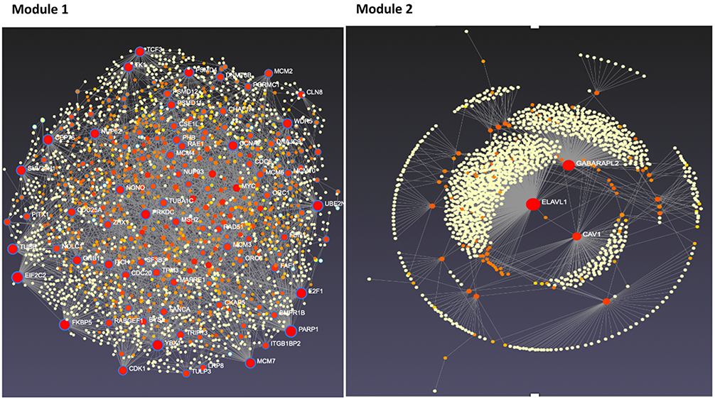
Figure 4: Tissue-specific protein-protein interaction network for modules 1 and 2 candidate genes.
The Trending With Impact series highlights Oncotarget publications attracting higher visibility among readers around the world online, in the news, and on social media—beyond normal readership levels. Look for future science news about the latest trending publications here, and at Oncotarget.com.
Listen to an audio version of this article
—
Hormones can cause tumor growth in some subtypes of breast cancer. Endocrine therapy, also known as hormone therapy, is a type of cancer treatment that removes or blocks the hormones which fuel breast cancer growth. This treatment is often given as adjuvant therapy after breast cancer surgery to lower the risk of cancer reoccurrence. In some cases, endocrine therapy may be used as a first-line treatment for hormone receptor-positive breast cancers, such as estrogen receptor-positive (ER-positive) breast cancer. However, ER-positive tumors frequently become unresponsive to endocrine therapy, and tumor regrowth can occur after treatment. The underlying causes of endocrine resistance are mostly undetermined.
“Endocrine therapies have been successful at improving cancer outcomes; however, the development of endocrine resistance, or resistance to inhibition of ER actions, remains a roadblock in breast cancer treatment.”
“In this study, we explored the dynamic behavior of the entire gene population to identify novel genes that play fundamental roles in the development and progression of endocrine-resistant breast cancer.”
Pipeline analysis in biology is a method of studying and analyzing a group of genes or proteins in order to understand their structure and function. The pipeline can be used to determine gene dynamics, clusters, similarities, and networks. In this case, the researchers used it to understand how endocrine resistance develops over time.
“The pipeline provides three main functions. First, statistical hypothesis testing determines a set of dynamic response genes (DRGs) that exhibit significant changes over time. Next, these DRGs are clustered into gene response modules (GRMs), sets of DRGs with similar time course expression patterns. Finally, the GRMs associations and regulatory effect are analyzed as a gene regulatory network using ordinary differential equations.”
The Study
To begin this study, the researchers first aimed to select a cell-based model that represents endocrine resistance in patients as closely as possible. They gathered data from breast cancer patients who were either resistant or sensitive to endocrine therapies and compared them with publicly available gene expression data. Results showed that the LTED MCF7 cell model displayed similar endocrine resistance to patient tumor data.
Next, the researchers observed the development of endocrine therapy resistance in the LTED MCF7 cell model, as well as the changes in gene expression over time. This data was collected and used to develop a mathematical model of gene expression dynamics during endocrine therapy resistance development. After statistical and computational pipeline analysis, the team identified a group of 254 genes whose time course expression significantly changed during the development of endocrine therapy resistance. They then aimed to validate their findings and used multiple bioinformatics approaches to narrow down this group of candidate genes.
“To further refine the genes common to endocrine resistance development and progression, we utilized several bioinformatic approaches designated to rank and prioritize the 254 common genes.”
The Results
Candidate genes were narrowed down to a novel group of 34 genes whose time course expression most significantly changed during LTED MCF7 cell modeling of endocrine-resistant breast cancer development. In addition, microarray analysis also showed that a subset of these genes was differentially expressed in triple-negative breast cancer (TNBC). This suggests that there may be shared genetic mechanisms between endocrine-resistant breast cancer and TNBC.
“As these two subtypes of breast cancer are the most fatal breast cancers with no known effective therapeutic approaches available to date, research on underlying genetic factors is of great importance.”
Conclusion
Their analysis led to the identification of a novel group of 34 genes that may play a role in endocrine resistance. Interestingly, some of these genes were also differentially expressed in TNBC. These findings could potentially lead to the development of new therapeutic strategies to overcome endocrine therapy resistance in some of the most difficult to treat and fatal breast cancers.
“Our analysis identified novel candidate genes with potential significance in endocrine-resistant breast cancer as well as TNBC, which opens new doors for designing novel therapeutic approaches for endocrine-resistant breast cancer and TNBC.”
Click hereto read the full research paper published by Oncotarget.
Oncotarget is an open-access journal that publishes primarily oncology-focused research papers in a continuous publishing format. These papers are available at no cost to readers on Oncotarget.com. Open-access journals have the power to benefit humanity from the inside out by rapidly disseminating information that may be freely shared with researchers, colleagues, family, and friends around the world.
In a new study, researchers investigated the role of the (pro)renin receptor in endometrial cancer cell growth.
Listen to an audio version of this article
In the United States and worldwide, the incidence and mortality rates of endometrial cancer among women have been increasing in recent years. While endometrial cancer is highly treatable, the primary treatment is a surgical hysterectomy. Hysterectomies can have serious side effects and painful personal consequences.
The ATPase H(+)-transporting lysosomal accessory protein 2 (ATP6AP2) gene encodes the (P)RR, and the terms can be used interchangeably. The (P)RR is a membrane protein that plays a key role in activating the renin-angiotensin system (RAS). It is widely expressed in various tissues and organs, such as the kidney, heart, lung, and endometrium. In endometrial cancer, the (P)RR has been shown to be overexpressed in cancerous tissue compared to normal endometrium tissue. Expression levels of this receptor are associated with endometrial cancer progression and poor prognosis. However, the precise role of the (P)RR in endometrial cancer has remained largely unknown.
In this in vitro analysis, the researchers first conducted a proteomic screening of the ATP6AP2 protein and mRNA expression in three endometrial cancer cell lines: Ishikawa, AN3CA and HEC-1-A. To silence (P)RR expression in each of the three cell lines, the team employed an siRNA-mediated knockdown of ATP6AP2. Next, they used an xCELLigence RTCA DP instrument that measures cell invasion and migration to evaluate the impact of (P)RR knockdown on cellular proliferation. They then used a resazurin assay to examine the effects of (P)RR knockdown on cancer cell viability.
A proteomic screening was also carried out to explore potential pathways (P)RR is involved in in the physiology of endometrial cancer. In addition, the enzyme-linked immunosorbent assay (ELISA) was used to measure circulating soluble prorenin receptor (s(P)RR) levels in the endometrial cancer cell lines (before and after the knockdown of (P)RR expression) and in plasma and uterine fluid samples donated by endometrial cancer patients.
The Results
This study was the first to report the mRNA and protein expression of (P)RR in three endometrial epithelial cancer cell lines. The results showed that the (P)RR was critical for endometrial cancer cell growth—contributing both to its cell viability and proliferative capacity. However, the data confirmed their previous observations that (P)RR mRNA and protein levels do not correlate with tumor grade in primary endometrial tumor samples. The researchers stated that the (P)RR’s contribution to endometrial cancer progression is likely mediated through proteins reduced after (P)RR expression knockdown, such as MGA, SLC4A7, SLC7A11, or DHRS2.
“Notably, (P)RR mRNA and protein levels were independent of tumour grade, with the highest expression detected in Ishikawa cells (grade 1), followed by AN3CA cells (grade 3) and finally HEC-1-A cells (grade 2).
They also observed that s(P)RR levels in their plasma samples were significantly higher in patients with endometrial cancer than in age-matched controls. Intriguingly, as cancer grade increased, so did s(P)RR levels. This indicated that s(P)RR may be a viable predictive or diagnostic marker for patients with endometrial cancer.
“Our data confirms that the (P)RR is important for endometrial cancer development, contributing to both its viability and proliferative capacity. Moreover, our quantitative proteomics approach uncovered several putative protein interactions and pathways that rely on (P)RR for disease progression and may represent novel therapeutic targets in the treatment of endometrial cancer. Finally, we contend that circulating s(P)RR levels may have substantial potential as a novel biomarker for cancer diagnosis and prognosis.”
Conclusion
This study sheds new light on the role of the (P)RR in endometrial cancer. The researchers suggest that future studies should aim to vet their findings in endometrial cancer patients.
“Collectively, our data indicate that targeting the (P)RR by an siRNA approach (such as in this study) or with an alternative anti-(P)RR monoclonal antibody approach currently being explored by Wang et al. [29] may be a viable therapeutic strategy against endometrial cancer.”
Click hereto read the full research paper published by Oncotarget.
Oncotarget is an open-access journal that publishes primarily oncology-focused research papers in a continuous publishing format. These papers are available at no cost to readers on Oncotarget.com. Open-access journals have the power to benefit humanity from the inside out by rapidly disseminating information that may be freely shared with researchers, colleagues, family, and friends around the world.