Researchers analyzed various publicly available datasets and identified two RNA modification regulatory proteins that are not only overexpressed in melanoma, but necessary for melanoma growth.
Malignant melanoma under the microscope.
The Trending with Impact series highlights Oncotarget publications attracting higher visibility among readers around the world online, in the news, and on social media—beyond normal readership levels. Look for future science news about the latest trending publications here, and at Oncotarget.com.
—
Listen to an audio version of this article
After melanoma of the skin metastasizes, it commonly becomes very difficult for doctors to treat. This is due to melanoma’s problematic tendency to acquire resistance to therapeutic interventions. Despite the development of many new targeted interventions and immunotherapies, five-year survival rates for patients with melanoma continue to be less than 10% for patients with lymph node metastasis and less than 5% for patients with distant metastasis.
“As the number of potential therapeutic DNA targets dwindle, many researchers are turning to RNA to tackle the problem.”
“Many studies have shown that these RNA modifications play crucial role in melanoma growth and metastasis [58, 59]. They are also involved in drug resistance mechanism.”
The Study
“Since RNA is a key molecule that drives every cellular process, their deregulation is present in nearly all human disease and play a causative role.”
The researchers explain that alterations among RNAs may arise due to altered activity or expression of the enzymes/proteins which are involved in the modification process. In this study, the team used multiple publicly available bioinformatics platforms to, first, analyze RNA alterations in melanoma samples, and then, to comprehensively analyze RNA modification regulatory proteins among melanoma samples. The publicly available datasets included: The Cancer Genome Atlas, The Human Protein Atlas, Oncomine, and the UALCAN database.
“Our study started with the analysis of various genetic alterations (amplifications, mutations/deletion) as well as RNA overexpression of these RNA modification regulatory proteins in The Cancer Genome Atlas melanoma database.”
Based on their analyses of these databases, reverse transcription quantitative PCR, soft-agar assays, validation by shRNA-mediated knockdown, and statistical analysis, the team identified what they believe are the most relevant RNA modifying proteins that play a crucial role in the development of melanoma. They found that METTL4 and DNMT3A RNA-modifying enzymes/proteins are both necessary for melanoma growth and overexpressed in melanoma.
“Based on this we infer that the upregulated expression of RNA modification regulatory proteins METTL4 and DNMT3A play a key role in melanoma initiation or progression.”
Conclusion
The researchers explained that their studies served the duel purpose of improving their understanding of novel pathways that cause melanoma to become untreatable, and also paving the way “to develop new, effective and sustainable therapeutic tools for optimal drug selection and treatment.”
“Additional future studies are needed to fully determine the role of these RNA modification regulatory proteins in melanoma tumor growth and progression (e.g., metastasis).”
Click here to read the full scientific study, published by Oncotarget
—
Oncotarget is a unique platform designed to house scientific studies in a journal format that is available for anyone to read—without a paywall making access more difficult. This means information that has the potential to benefit our societies from the inside out can be shared with friends, neighbors, colleagues, and other researchers, far and wide.
In this trending study, the association between IGF/CTP composite scores, overall survival, and progression-free survival of hepatocellular carcinoma patients treated with sorafenib was investigated.
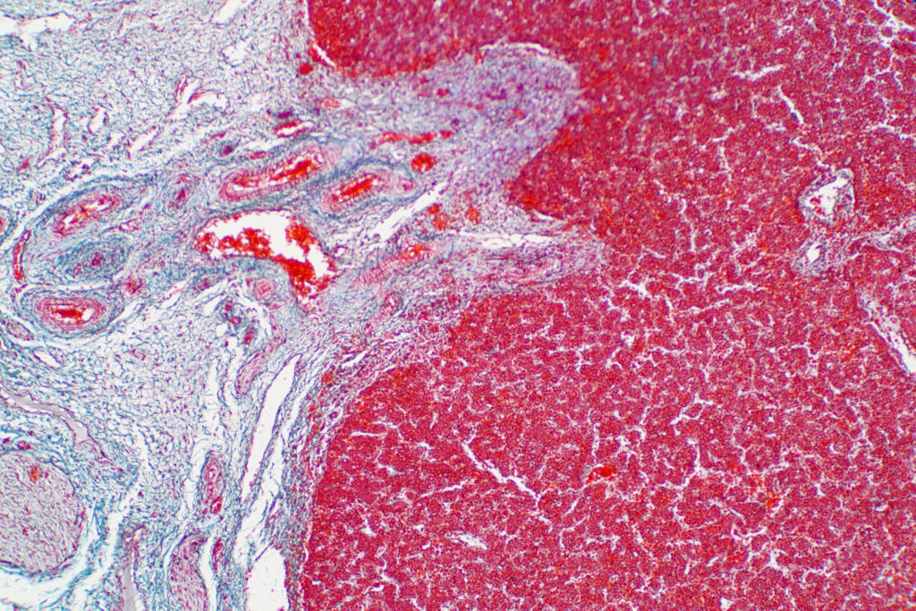
Human liver tissue under the microscope view.
The Trending with Impact series highlights Oncotarget publications attracting higher visibility among readers around the world online, in the news, and on social media—beyond normal readership levels. Look for future science news about the latest trending publications here, and at Oncotarget.com.
—
Listen to an audio version of this article
Sorafenib was the first systemic therapy approved to treat Child-Turcotte-Pugh (CTP) class A patients with advanced hepatocellular carcinoma (HCC). However, there are no biomarkers known to predict survival, treatment outcomes, or to guide this HCC systemic therapy. The insulin-like growth factor 1 Child-Turcotte-Pugh (IGF-1/CTP) composite score has emerged as a hepatic reserve assessment tool—and potential prognostic biomarker.
“Accurate assessment of the functional hepatic reserve is important to the prognostic and treatment prediction for patients with liver disease [1, 2].”
The majority of circulating insulin–like growth factor (IGF) is synthesized and secreted by the liver, and levels of IGF dramatically decrease in chronic liver disease and HCC. IGF can be a helpful tool to determine the prognosis of patients with advanced HCC while undergoing treatment with sorafenib. Researchers also use the Child-Turcotte-Pugh (CTP) qualitative scoring system to assess severity of liver cirrhosis, hepatic reserve, guide treatment decisions, and to stratify patients with HCC into three groups (A, B, and C). CTP class A has a better prognosis compared to classes B and C.
“Assessing liver reserve in HCC is of a great value as a tool for stratification of patients in clinical trials as well as to predict HCC outcome and guide therapy decisions in routine practice [28].”
In the researchers’ prospective study, 171 patients with HCC from the University of Texas MD Anderson Cancer Center were screened and included in this study. Of the patients, 116 were classified in CTP group A. Patient IGF/CTP scores were calculated and the researchers used the Kaplan-Meier method and log-rank test to estimate and compare the time-to-event outcomes between patient subgroups. Based on CTP and the IGF/CTP scores, researchers reclassified group A patients into AA and AB risk groups, which differed significantly in terms of OS and PFS. The researchers followed up with all patients in the study until disease progression or death. Unfortunately, during the follow-up period, 100 patients passed away.
“After IGF/CTP scoring, 87 of 116 CTP class A patients were reclassified as IGF/CTP-A (AA) and 29 patients were reclassified as IGF/CTP-B (AB) (Supplementary Table 1).”
Results & Conclusion
“Our study is the first prospective validation of the IGF/CTP scoring system association with the outcomes among patients with HCC treated with sorafenib.”
This study supported the researchers’ hypothesis that the IGF/CTP score is capable of further distinguishing and refining CTP class A patients. However, for CTP class A patients, due to limited power of the study, researchers were unable to meet the threshold for statistical significance for the OS and PFS durations of the reclassified groups AA and AB.
“Although our study was not powered to determine the predictive value of the IGF/CTP score in regard to median OS and PFS durations in CTP class A patients treated with sorafenib, our subset analyses of OS and PFS at different timepoints were statistically significant and, if independently validated, could change the standard approach to assessing hepatic reserve in patients with HCC.”
Click here to read the full scientific study, published by Oncotarget.
—
Oncotarget is a unique platform designed to house scientific studies in a journal format that is available for anyone to read—without a paywall making access more difficult. This means information that has the potential to benefit our societies from the inside out can be shared with friends, neighbors, colleagues, and other researchers, far and wide.
Oncotarget is a proud participant of the AACR Annual Meeting 2021 #AACR21
Researchers examined midostaurin resistance or sensitivity in a cohort of patients with acute myeloid leukemia.
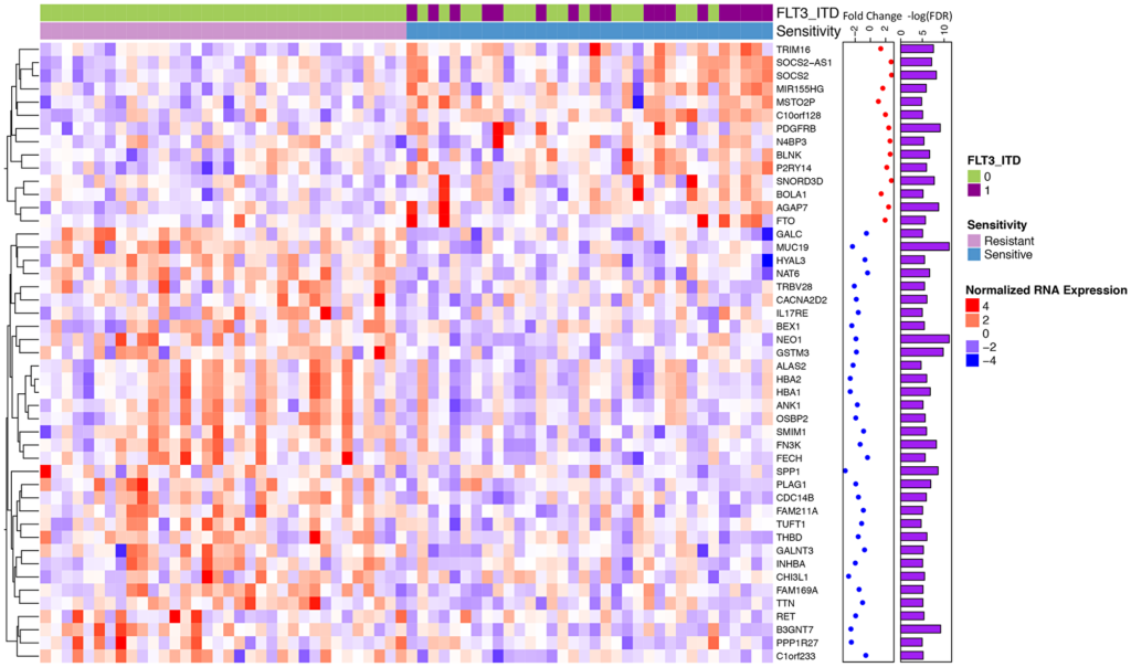
Figure 2:Differential gene expression for midostaurin sensitive vs. resistant samples identifies a unique signature.
The Trending with Impact series highlights Oncotarget publications attracting higher visibility among readers around the world online, in the news, and on social media—beyond normal readership levels. Look for future science news about the latest trending publications here, and at Oncotarget.com.
—
Listen to an audio version of this article
Acute myeloid leukemia (AML) is a heterogeneous malignancy that most commonly affects older adults, 60 years of age and older. NPM1, DNMT3A, and FLT3 are the most common genomic alterations found within this disease. In about 30% of AML patients, FLT3 is mutated. Midostaurin was the first FDA approved FLT3 inhibitor for AML. While Midostaurin has a successful overall survival benefit, both primary and secondary resistanceremains common.
“A subtype of AML, classified by the presence of a FLT3-Internal Tandem Duplication (ITD) mutation, tends to have a worse prognosis with early relapse and death [5].”
In order to understand the impact that different genomic alterations have on midostaurin response, 214 patients were functionally assessed with midostaurin and their FLT3 status was annotated. Of these patients, the researcher identified 193 primary and 21 relapse AML samples from the Beat AML publicly available dataset. Risk groups within the cohort were as follows: 73 samples were favorable risk, 59 samples were intermediate, and 68 were adverse. The median age of patients in the cohort was 61, with 52% male and 48% female.
“We hypothesized that there are additional genomic alterations and gene expression changes outside of FLT3-ITD mutations that can influence AML sample resistance or sensitivity to midostaurin and aimed to further characterize these factors.”
Drug sensitivity screening, RNA sequencing/expression analysis, custom gene panel (GeneTrails) sequencing and variant detection, exome sequencing and variant detection, internal FLT3-ITD and NPM1 mutation detection, derivation of FLT3-ITD and NPM1 consensus calls, ex vivo functional drug screens, and statistical analysis were the methods used to observe the impact of genomic alterations on midostaurin response.
“Our research explored the multi-targeted nature of midostaurin and suggested a number of molecular mutational patterns that correlated with midostaurin drug sensitivity and resistance in both FLT3-ITD mutated and FLT3-ITD wild-type AML patient samples.”
Results
The researchers observed specific point mutations and gene expression patterns that they believe explain why there is a range of responses to midostaurin treatment. In the FLT3-ITD positive cohort, increased expression of the oncogene RGL4 (and regulator of the Ras-Raf-MEK-ERK cascade) correlated with poorer midostaurin response. In the FLT3-ITD negative cohort, KRAS mutations correlated with a poorer midostaurin response.
“We also observed that 16 / 34 of the most sensitive samples did not harbor a FLT3 mutation and a majority of differentially expressed genes were independent of FLT3 status.”
Conclusion
The authors point out that additional research studies will be needed given that their sample cohort was relatively small. They also note that since there are multiple FLT3 inhibitors available, it is important to understand the sensitivity mechanisms of each intervention in order to better personalize therapy for chemo-refractory or relapsed AML patients.
“Overall, we identify genomic alterations that correlate with midostaurin response independent of FLT3-ITD status, propose that Ras-Raf-MEK-ERK inhibition in combination therapy could limit resistance to midostaurin, and suggest that within the overall AML population there may be therapeutic benefit of midostaurin in patients with certain expression profiles.”
Click here to read the full scientific study, published in Oncotarget.
—
Oncotarget is a unique platform designed to house scientific studies in a journal format that is available for anyone to read—without a paywall making access more difficult. This means information that has the potential to benefit our societies from the inside out can be shared with friends, neighbors, colleagues, and other researchers, far and wide.
Researchers compared vitamin D binding protein expression in papillary thyroid cancer tissues among Filipino American and European American patients.
3D rendered medically accurate illustration of thyroid cancer.
The Trending with Impact series highlights Oncotarget publications attracting higher visibility among readers around the world online, in the news, and on social media—beyond normal readership levels. Look for future science news about the latest trending publications here, and at Oncotarget.com.
—
On the basis of ethnicity, different gene variants of the vitamin D binding protein (DBP) are expressed among different populations of people around the world. Little is known about this highly polymorphic protein, though, researchers do know that DBP functions dependently and independently of vitamin D. Many previous research studies have examined the vitamin D-dependent correlation between DBP and cancer, however, few studies have examined DBP’s functionality independent of vitamin D, especially regarding the role of DBP expression in thyroid cancer.
“A systemic review demonstrated that a large number of chronic diseases, including cancers, have been associated with DBP variants [29].”
“Although DBP is an essential protein with multifunctional properties, [28, 41–47], very few studies are available on its contribution to thyroid cancer oncogenesis.”
The Study
“Thyroid cancer incidence, recurrence, and death rates are higher among Filipino Americans than European Americans.”
To determine the correlation between differential DBP expression in tumor tissues and cancer staging among Filipino Americans and European Americans, the researchers gathered 200 archival papillary thyroid tissues; 100 from Filipino Americans and 100 from European Americans. They used immunohistochemistry to assay DBP expression in each sample and then analyzed the data with confocal microscopy.
“Since DBP gene variants showed differential expression across ethnicities [25, 40, 48, 49], DBP level in the tumor microenvironment may implicate the difference in TC [thyroid cancer] prognosis between Filipino and European Americans.”
First, the team evaluated whether or not there was any relationship between their DBP staining results and age, gender, or body mass index of the patients. They found no correlation between DBP levels and any of these variables, in either ethnicity. The researchers then analyzed the immunohistochemistry DBP staining results by ethnicity. They found moderate to strong intensity DBP staining across the European American cancer tissues and significantly low to no DBP staining in the Filipino American cancer tissues. The researchers also determined an inverse relationship between DBP expression and cancer stage—the lower the DBP expression, the poorer the prognosis.
“These data implied that DBP’s presence might play protective roles in cancer progression in European Americans compared to Filipino Americans, supporting the aggressive phenotype observed in Filipino Americans.”
Next, to observe the effects on cell migration and proliferation, DBP knockdown and overexpression (almost 90%) was achieved in the papillary thyroid tumor cells. The researchers demonstrated increased cancer cell proliferation and migration after the knockdown of the DBP gene. When the researchers overexpressed DBP, they observed a significant reduction in papillary thyroid cancer cell proliferation and migration.
Conclusion
“In conclusion, we demonstrate that the presence or absence of DBP inversely correlates to thyroid cancer staging in two ethnicities.”
The researchers note that while this study demonstrated low vitamin D binding protein expression in the advanced thyroid tumors of Filipino Americans, they acknowledge the need to determine the progressive loss of DBP throughout the stages of thyroid cancer.
“A future study is underway to determine the DBP regulation and its downstream pathways to elucidate strategies to eliminate the observed thyroid cancer health disparities.”
Click here to read the full scientific study, published in Oncotarget.
—
Oncotarget is a unique platform designed to house scientific studies in a journal format that is available for anyone to read—without a paywall making access more difficult. This means information that has the potential to benefit our societies from the inside out can be shared with friends, neighbors, colleagues, and other researchers, far and wide.
Oncotarget, exhibited by its publisher Impact Journals, will be participating virtually at the AACR Annual Meeting this year, from April 10-15 and May 17-21, 2021.
Oncotarget participating in the Annual AACR Meeting 2021 #AACR
Listen to an audio version of this article
The American Association for Cancer Research (AACR) organizes an annual meeting program covering some of the most recent discoveries in cancer research. The conference aims to highlight work from the best minds in research and medicine from institutions all over the world. Oncotarget, exhibited by its publisher Impact Journals, will be participating virtually at the AACR Annual Meeting this year.
As of June 2020, Scopus released their latest 2019 Journal Rankings on Oncology. Oncotarget is among their highest rated (Q1) journals and ranked number one in total citations in oncology. The journal has published outstanding papers and reviews by authors including Bert Vogelstein, Peter K. Vogt, Pier Paolo Pandolfi, Arnold J. Levine, Brian Druker, and Carol Prives. Founding Oncotarget Editorial Board members include Nobel Laureates Andrew V. Schally and Gregg L. Semenza; Lasker Award recipients Alexander Varshavsky, Brian J. Druker, and Gregg L. Semenza; and 16 members of the US National Academy of Sciences. Oncotarget is indexed and archived in PubMed, PubMed Central, Scopus, EMBASE, and META (Chan Zuckerberg Initiative) .
The 2021 AACR conference, a two-week online event, will take place from April 10-15 and May 17-21, 2021. Topics include population science and prevention, cancer biology, translational and clinical studies, survivorship, and advocacy.
In 2019, Oncotarget participated in the AACR Annual Meeting at the Georgia World Congress Center in Atlanta, Georgia, USA, and “AACR-NCI-EORTC International Conference on Molecular Targets and Cancer Therapeutics,” at the Hynes Convention Center in Boston, Massachusetts, USA. The total registration count from the 2019 AACR Annual Meeting was over 21,000—nearly 16,000 of which were scientific attendees from all over the world. Click here to view photos from Oncotarget’s participation in the 2019 AACR Annual Meeting.
Follow the Oncotarget Twitter account (@Oncotarget) for live updates about the conference using the #AACR21 hashtag.
—
Oncotarget is a unique platform designed to house scientific studies in a journal format that is available for anyone to read—without a paywall making access more difficult. This means information that has the potential to benefit our societies from the inside out can be shared with friends, neighbors, colleagues, and other researchers, far and wide.
Researchers conducted a 2021 study to better characterize phyllodes tumors and other breast fibroepithelial lesions in order to improve diagnosis and treatment for patients.
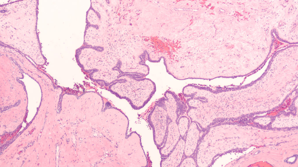
Photomicrograph showing histology of a benign phyllodes tumor of the breast, from sections of an excision specimen (lumpectomy).
The Trending with Impact series highlights Oncotarget publications attracting higher visibility among readers around the world online, in the news, and on social media—beyond normal readership levels. Look for future science news and articles about the latest trending publications here, and at Oncotarget.com.
—
Listen to an audio version of this article
Thankfully, around 80% of lumps found in human breasts turn out to be benign, or indolent, fibroadenoma (FAD). FADs fall into a category of breast fibroepithelial lesions (FELs), which include many heterogeneous pathological tumors, ranging from benign FADs to rare and potentially aggressive phyllodes tumors (PTs). After examination by a physician, these FELs may be diagnosed as either benign, borderline, or malignant.
“The current grading system remains unreliable in differentiating these tumors due to histological heterogeneity and lack of appropriate markers to monitor the sudden and unpredictable malignant transformation of PTs.”
The Study
To begin identifying the differentially expressed genes and proteins among FADs and PTs in benign, borderline, and malignant states, the researchers conducted quantitative global proteomics on Formalin-Fixed Paraffin-Embedded (FFPE) tissue sections. They conducted a principal component analysis of the protein expression matrix to identify the overlapping proteomic profiles among FELs.
“Interestingly, we observed FADs and benign PTs clustered together compared to borderline and malignant ones, albeit with overlapping protein expression profiles.”
When FADs were compared with benign PTs, the researchers identified 32 proteins in FAD that were differentially regulated. The researchers elucidated many important distinctions between benign, borderline, and malignant FADs and PTs, and identified at least three potential prognostic markers that may aid in patient diagnosis and treatment. The progression of PTs from borderline to malignant and their mechanistic framework was clearly explained by the researchers in this study.
“The presence of extensive ECM proteins and EMT markers led us to hypothesize a model of deposition and degradation of these proteins thus triggering ECM remodeling and EMT acquisition in borderline PTs leading to its malignant state. Enrichment of platelet degranulation factors in malignant PT indicates active angiogenesis during this transformation.”
The Study
“Herein, our initial findings suggest that MUCL1, HTRA1, and VEGFD can be used as potential proteomic markers that could augment existing diagnosis, and help in monitoring the progression of the disease.”
Additional characterization of FELs using different omics platforms was recommended by the researchers to help better understand and manage the dynamics of PTs and malignant breast tumors.
“The present work shed light on a brief mechanistic framework of PTs aggressive nature and present potential biomarkers to differentiate overlapping FELs that would be of practical utility in augmenting existing diagnosis and disease management for this rare tumor.”
Click here to read the full scientific study, published in Oncotarget.
—
Oncotarget is a unique platform designed to house scientific studies in a journal format that is available for anyone to read—without a paywall making access more difficult. This means information that has the potential to benefit our societies from the inside out can be shared with friends, neighbors, colleagues, and other researchers, far and wide.
Young people living in the Andes Mountains are disproportionately affected by hepatocellular carcinoma compared to other youth around the world. Researchers conducted a study to better understand the cause.
Peru. View of the Urubamba River through the Aguascalientes Village.
The Trending with Impact series highlights Oncotarget publications attracting higher visibility among readers around the world online, in the news, and on social media—beyond normal readership levels. Look for future science news and articles about the latest trending publications here, and atOncotarget.com.
—
Listen to an audio version of this article
Andean people live in sparsely populated regions in the Andes Mountains of South America. It is the longest mountain range in the world; spanning seven countries from southern Peru to southern Argentina. Due to the high elevations (averaging 13,000 feet; peaking at 22,834 feet), these areas are known for such low oxygen levels that Andean people have adapted physiologically to the extreme conditions.
Around the world, hepatocellular carcinoma (HCC) is the main form of primary liver cancer and commonly affects older patients after they have had prolonged liver disease. However, among Andean people, half of the total patients who develop HCC are adolescents and young adults. Researchers—from Sorbonne Université, Institut Pasteur, Université de Rennes, and Université de Toulouse in France, and the Instituto Nacional de Enfermedades Neoplásicas in Peru—conducted a study to better understand HCC in Andean people.
“To deepen our understanding of the molecular determinants of the disease in this population, we conducted an integrative analysis of gene expression and DNA methylation in HCC developed by 74 Peruvian patients, including 39 adolescents and young adults.”
The Study
“The 74 Peruvian patients with HCC included in the present study carried mitochondrial DNA (mtDNA) haplotypes of the four ancestral lineages (A–D) shared by Indigenous American populations (Figure 1A and Table 1) [23].”
The researchers retrospectively conducted transcriptome profiling of patient samples from 74 Peruvian patients with HCC. They compared gene expression data (after batch-effect removal) and found that Peruvian HCC is characterized as a distinct molecular subtype. This, now referred to as the “Amerind signature,” identifies Peruvian HCC as a distinct phenotypic cluster.
“A 961 gene signature was defined (hereinafter referred to as “Amerind signature”), of which 806 were upregulated and 155 downregulated in Peruvian HCC (Figure 3A and Supplementary Table 4).”
Methylome profiling was also conducted by the researchers to show the dynamics of DNA methylation marks, which revealed that Peruvian HCC is associated with a genome-wide hypermethylation pattern. They explain that DNA hydroxymethylation also represents a relevant epigenetic mark in Peruvian HCC. In addition, the researchers found evidence that Peruvian HCC tumor cells have a weaker retinoid signaling signature, which opens the door to potential therapeutic targets.
“The genomic analysis of Peruvian HCC evidenced a weaker retinoid signaling signature in tumor cells, which could pinpoint novel targets and drugs for anticancer targeted therapy (Figure 1C and Supplementary Table 1) [45]. We hypothesized that this weaker retinoid signaling could be responsible for the increased proliferation; hence, the pharmacological response to RA should antagonize this process.”
Conclusion
After comparing this sample of patients with Peruvian HCC with other HCC tumors from other countries around the world, molecular divergence in Peruvian HCC was demonstrated by showing “hierarchical clustering relying on a large and meaningful gene expression signature.” The researchers do not yet know if these differences are due to external/geographic or genomic factors.
“Whether this molecular phenotype is due to anthropological specificities embedded in genome architecture, to extrinsic etiological cues, or to subtle interplays between both components remains to be ascertained.”
With this being said, the researchers believe that this study stresses the need to carefully consider the potentially prominent roles of human genomic architecture and biogeography when it comes to cancer and underreported minorities and Indigenous patients, especially in low- and middle-income countries. They are forthcoming about limitations in their study and mention having analyzed a fairly small sized cohort. Importantly, the findings from this study create a case for developing therapeutics that are tailored to this new molecular subtype of HCC.
“The present study establishes a foundation for the dissection of the functional importance of RA-mediated epigenetic control in HCC and therapeutics tailored to patients with Indigenous American ancestry.”
Click here to read the full scientific study, published in Oncotarget.
—
Oncotarget is a unique platform designed to house scientific studies in a journal format that is available for anyone to read—without a paywall making access more difficult. This means information that has the potential to benefit our societies from the inside out can be shared with friends, neighbors, colleagues and other researchers, far and wide.
Researchers use a computer simulated modeling system to highlight the strengths and weaknesses of two ALK inhibitors.
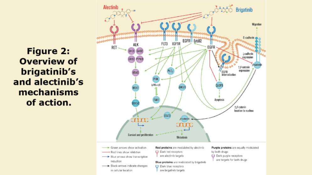
Figure 2: Overview of brigatinib’s and alectinib’s mechanisms of action.
The Trending with Impact series highlights Oncotarget publications attracting higher visibility among readers around the world online, in the news, and on social media—beyond normal readership levels. Look for future science news and articles about the latest trending publications here, and at Oncotarget.com.
—
Listen to an audio version of this article
Despite many therapeutic advances over the years, over half of patients with lung cancer die within one year of diagnosis. Non-small cell lung cancer (NSCLC) comprises 85% of all lung cancer, and around 3–7% of patients with NSCLC present with a rearranged ALK gene (ALK+). This abnormality produces aberrant ALK protein cell signaling pathway activity and causes cancer cells to grow and metastasize. ALK+ NSCLC patients often develop drug resistance to available ALK inhibitor drugs.
“Consequently, it is of the upmost importance to adequately use the currently available treatments in the correct order to maximize the life span of NSCLC patients.”
The researchers first began their study by characterizing the pathophysiology of ALK+ NSCLC after completing a detailed review of review papers published in PubMed between 2013 and 2018.
“To carefully characterize the pathophysiology of ALK+ NSCLC, we conducted an extensive and detailed full-length review of relevant review articles over the last 5 years in the PubMed database (from December 3rd 2013 to December 3rd 2018)[…]”
Next, to compare the strengths and weaknesses of two second generation ALK inhibitor drugs, brigatinib and alectinib, the researchers used a computer simulated modeling system—in silico. They explain that an in silico method of study can be highly useful when analyzing drug characteristics and predicting the biochemical characteristics and drug mechanisms of action.
“Overall, these systems can be employed for the exploration of anticancer drug mechanisms of action and their efficacy in specific patient profiles.”
The in silico system they used is called a Therapeutic Performance Mapping System (TPMS) and is based on artificial intelligence and pattern recognition models. This TPMS system was “trained” by the researchers in this study and given up-to-date biological and clinical data to input into its configuration. The mathematical models used to obtain the ALK inhibitors’ mechanisms of action were generated following the same methodology as described in this study.
“This methodology integrates available biological, pharmacological and medical information to generate mathematical models that simulate the mechanisms of action of drugs in a pathophysiological human context (Figure 4).”
To detect and explain the biological relationships that occur, the team used two distinct modeling methods: artificial neural networks and sampling-based methods. They applied Sobol sensibility analysis over the TPMS mathematical models in order to account for the impact of any noise affecting the final mechanisms of action. The researchers also performed drug-(patho)physiology motive relation finding and evaluated the impact of potential resistances and drug interferences over the mechanisms of action.
Results & Conclusion
“According to the current knowledge and the data herein presented, brigatinib might be more prone to present relevant metabolic and mechanistic interactions with other drugs than alectinib, which might be a safer option in poly-treated patients.”
“Brigatinib appears to have a wider mechanism of action, presenting targets that potentially act more strongly in most of the ALK+ NSCLC pathophysiological pathways, including invasiveness to the CNS [central nervous system].”
“On the other side, alectinib-induced RET inhibition might contribute to reducing the tumour immune evasion mechanisms.”
The researchers found that both drugs are known to be well-tolerated and show similar efficacy for the treatment of ALK+ NSCLC in a first-line setting. However, they explain that the differences in their characteristics shown in this study might allow for administration in more targeted patient populations that might see benefits from either brigatinib or alectinib. This deeper classification may also help when considering potential safety concerns in specific patient subpopulations.
“Future clinical studies will be needed to confirm these findings. The used approach can be applied for the evaluation of other next-generation ALKi, even if not yet approved, or exploring other questions, such as optimal treatment sequence.”
Click here to read the full scientific study, published in Oncotarget.
—
Oncotarget is a unique platform designed to house scientific studies in a journal format that is available for anyone to read—without a paywall making access more difficult. This means information that has the potential to benefit our societies from the inside out can be shared with friends, neighbors, colleagues and other researchers, far and wide.
Authors of this review paper discuss the complex crosstalk between cancer stem cells and macrophages, and potential anti-cancer strategies for future studies.
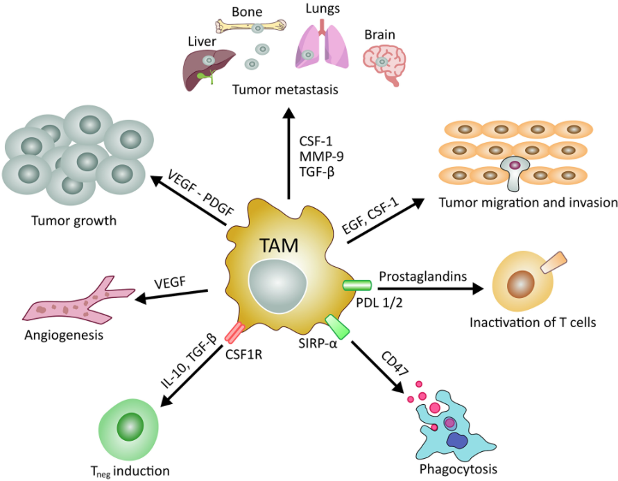
Figure 1: Main roles of tumor associated macrophages in cancer development and maintenance.
The Trending with Impact series highlights Oncotarget publications attracting higher visibility among readers around the world online, in the news, and on social media—beyond normal readership levels. Look for future science news and articles about the latest trending publications here, and at Oncotarget.com.
“The aim of this review is to define the complex crosstalk between these two cell types and to highlight potential future anti-cancer strategies,” Dr. Beatrice Aramini said, a thoracic surgeon and scientist from the University Hospital of Modena Reggio Emilia.
There have been numerous studies published over recent decades in an effort to understand the molecular mediators of cancer stem cells (CSCs) and tumor associated macrophages (TAMs). Several studies have contributed to bringing light to some of the complex crosstalk that occurs between these two cell types and within the tumor microenvironment. The authors of this review paper reference hundreds of studies and offer a thorough audit and analysis of the current state of this research.
About the Writers of the Review Article
“I mainly focus on lung cancer,” Dr. Aramini said. “I started this project about cancer stem cells in lung cancer since 2017, at University Hospital of Modena Reggio Emilia, joining the laboratory of cell therapies directed by Professor Massimo Dominici with the chief of medical oncology at University Hospital in Modena.”
Dr. Valentina Masciale, co-author, is a research fellow at the University Hospital of Modena Reggio Emilia. Dr. Masciale’s professional experience began by studying missing stroma cells. She then studied stem cells in regenerative medicine and currently she is working with Dr. Aramini on a project focused on lung cancer stem cells. The paper they wrote was revised and approved by seven other contributing authors.
“In this review, we describe the importance of cancer stem cells as the key drivers of cancer initiation and progression due to their unlimited cell renewal capacity and their ability to induce tumor formation,” Dr. Aramini said.
Introduction to Cancer Stem Cells
“Cancer stem cells (CSCs) constitute a cancer cell subpopulation similar to the other stem cell types in terms of self-renewal and multilineage differentiation potential but drive tumor development besides heterogeneity and dissemination of cancer cells [1–9].”
In 1997, Bonnet and Dick were the first researchers to report the existence of cancer stem cells in the tumor, in acute myeloid leukaemia. Since then, however, a standard marker to identify CSCs still has yet to be found.
“One of the main obstacles to proving the CSC model is the difficulty in identification and isolation of these cells [7, 33, 91].”
The authors explain that one of the problems with finding a marker such as this is that many markers found are not only able to detect CSCs, but they also detect non-tumor cells. This represents a major obstacle when developing new therapies to target CSCs only. The authors note that recently there have been several gene markers described by researchers for CSCs in different tumors, including brain, breast, blood, and lung.
“Indeed, there are currently no markers able to distinguish between stem cells and CSCs. Thus far, the best markers identified are those of onco-fetal stem cells, which are absent in adult organs and present in cancer cells [45–48].”
Theories About the Role of Cancer Stem Cells
This review refers to a few theories about the role of CSCs in cancer progression. One theory is based on the premise that tumor tissue is hierarchically organized into different types of cells, with the CSC subpopulation as the top of this hierarchy. In this theory, the other levels consist of additional differentiated tumor cells or cells with a limited proliferative potential. The “clonal evolution theory” hypothesizes that a rampant mutating cell is the catalyst for tumor progression.
“Peter Nowell was the first to describe the ‘clonal evolution theory,’ defining cancer as a complex process resulting from the development of a single out-of-control cell with multiple cell mutations that result in the progression of the tumor, which is kept viable through the selection of the most aggressive clones [89].”
Since the discovery of CSC plasticity and the possibility of switching from stem to non-stem cells, researchers have gained a more complex picture of the origin of tumor heterogeneity and more theories about the role of cancer stem cells in tumor progression.
“An opposing theory is based on the concept that CSCs are a group of cells endowed with a high self-renewal capacity that can set different phenotypes of tumorigenic cells [18, 88].”
Cancer Stem Cells and Macrophages
The researchers explain that macrophages are large specialized phagocytic cells that exist in tissues or at infection sites which act as part of the immune system. Arising from the bone marrow, macrophages perform multiple functions and roles in normal and tumor microenvironments, including pro-inflammatory activities and anti-inflammatory processes. Tumor-associated macrophages (TAM) comprise up to 50% of the tumor mass and have a close relationship with CSCs.
“The rising interest on these type of cells comes from recent study demonstrating that high number of tumor-associated macrophages correlate with the poor clinical prognosis in many solid tumors, including lung cancer, which is the field of our research group at the University Hospital of Modena,” Dr. Masciale said. “Another important aspect is the protective role of the tumor-associated macrophages play on tumors undergoing chemotherapy, which may impact the chemotherapy resistance and consequent tumor relapse.”
In recent studies, high numbers of TAMs in lung tumors, gastric cancer, and other cancer types, have been shown to correlate with a poor clinical prognosis. Macrophages are recruited to the tumor and, through crosstalk, provide protection to the tumor, contribute to immunosuppression in the tumor microenvironment and, eventually, drug resistance.
“The primary cause of failure in cancer treatment is the emergence of drug resistance that promotes the tumor spreading,” Dr. Masciale said.
“Cross-talk between CSCs and TAMs involves the recruitment of TAMs through vascularization and the release of chemokines by TAMs to preserve the quiescence of CSCs and modification of their antigens to escape from recruitment by immune cells.”
Future Perspectives
“Although most TAM-targeting strategies are in the pre-clinical stages, several factors used for TAMs depletion have already been tested in clinical trials [271, 272].”
Current efforts are underway to reprogram or inhibit the tumor-protective properties of tumor associated macrophages. Researchers are also investigating potential strategies to increase the efficacy of chemotherapy through nano-drug delivery to TAMs.
“Due to the significance of the tasks in which TAMs are involved, TAMs are increasingly becoming principal targets of novel therapeutic approaches, especially in the field of nanomedicine.”
The authors believe that targeting TAMs could trigger various reactions in the tumor, which are difficult to predict even given the individual variability from patient to patient. They also explain that targeting TAMs with CSCs offers another potential for treating different tumors to better control cancer progression and avoid tumor dissemination.
“In summary, generating new information about the interaction between TAMs and CSCs will be one of the most important challenges for the development of more effective targeted cancer therapies.”
Click here to read or watch an interview with the authors Behind the Study.
—
Oncotarget is a unique platform designed to house scientific studies in a journal format that is available for anyone to read—without a paywall making access more difficult. This means information that has the potential to benefit our societies from the inside out can be shared with friends, neighbors, colleagues and other researchers, far and wide.
Researchers in this study employed one of the few available murine cachexia models and validated its ability to be used in future studies of cancer-derived myocardial damage.
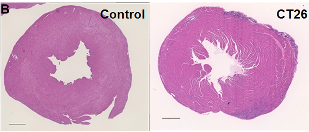
Part of Figure 2: Alterations in the myocardium of CT26-inoculated BALB/c mice.
The Trending with Impact series highlights Oncotarget publications attracting higher visibility among readers around the world online, in the news, and on social media—beyond normal readership levels. Look for future science news and articles about the latest trending publications here, and at Oncotarget.com.
—
Listen to an audio version of this article
Cachexia, a complex metabolic syndrome characterized in part by the loss of muscle mass, can account for up to 30% of all cancer-related deaths. Myocardial atrophy, or cardiac remodeling/degradation, is a phenotype of cachexia and a common cause of death.
“The causes of cancer-derived myocardial impairment might be the effects of cancer itself, background heart disease, and influence of cancer treatments; however, they have not been given much clinical importance, and specific treatment efforts are delayed [8].”
Researchers from Nara Medical University, Hanna Central Hospital, and Hoshida Minami Hospital in Nara and Osaka, Japan, and Nantong University in Jiangsu Province, China, note that while myocardial damage in cancer patients is known to be a cause of death, there are few murine cachexia models available to evaluate cancer-derived heart disorders. Thus, there is a need for further studies that may allow researchers to establish an intervention to prevent myocardial damage in cancer patients.
“In this study, we used the mouse cancer cachexia model that we previously established [14] to examine the status of cancer-derived myocardial impairment reported in literature, and validate our model for studying cancer-derived myocardial impairment.”
The Study
Some causes of cancer-derived myocardial impairment have been reported as cancer-induced cytokines, oxidative stress, depletion of antioxidants, and protein catabolism as a result of AKT/mTOR inhibition.
“Despite these advances in our understanding, the multifactorial mechanisms underlying cancer-derived myocardial impairment remain incompletely understood, necessitating further investigations to elucidate the molecular mechanisms and prevent myocardial damage in cancer patients.”
The researchers previously established a mouse cancer cachexia model. In this study, they aimed to validate their model by employing it in the examination of cancer-derived myocardial impairment that has been reported in previous literature. Their study enlisted the mouse model, CT26 colon cancer cell cultures, protein extraction, histological analysis, immunoblot analysis, enzyme-linked immunosorbent assay (ELISA), mitochondrial stress tests (Seahorse assay), glycolytic stress tests, and statistical analysis.
Conclusion
“In summary, our established mouse cachexia model showed various myocardial changes associated with cancer cachexia such as oxidative stress in the myocardium, energy metabolism, autophagy, and inflammatory cytokines.”
Results obtained by the researchers in this study using their mouse cachexia model are congruent with previously reported results about cancerous myocardial damage, and therefore provide reasonable evidence that it may be used in future studies.
“The established mouse cachexia model can therefore be considered useful for analyzing cancer-derived myocardial damage.”
Click here to read the full scientific study, published in Oncotarget.
—
Oncotarget is a unique platform designed to house scientific studies in a journal format that is available for anyone to read—without a paywall making access more difficult. This means information that has the potential to benefit our societies from the inside out can be shared with friends, neighbors, colleagues and other researchers, far and wide.